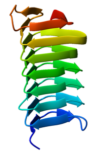
Monomeric, left-handed β-helix antifreeze protein from the spruce budworm Choristoneura fumiferana (PDB: 1M8N).Dimeric, right-handed β-helix antifreeze protein from the beetle Tenebrio molitor (PDB: 1EZG). Face-to-face association of β-helices.
A beta helix is a tandem protein repeat structure formed by the association of parallel beta sheet in a helical pattern with either two[1] or three[2] faces. The beta helix is a type of solenoid protein domain. The structure is stabilized by inter-strand hydrogen bonds, protein-protein interactions, and sometimes bound metal ions. Both left- and right-handed beta helices have been identified. These structures are distinct from jelly-roll folds, a different protein structure sometimes known as a "double-stranded beta helix".[3][4]
The first beta-helix was observed in the enzyme pectate lyase, which contains a seven-turn helix that reaches 34 Å (3.4 nm) long. The P22 phage tail spike protein, a component of the P22 bacteriophage, has 13 turns and in its assembled homotrimer is 200 Å (20 nm) in length. Its interior is close-packed with no central pore and contains both hydrophobic residues and charged residues neutralized by salt bridges.
Both pectate lyase and P22 tailspike protein contain right-handed helices; left-handed versions have been observed in enzymes such as UDP-N-acetylglucosamine acyltransferase and archaeal carbonic anhydrase.[5] Other proteins that contain beta helices include the antifreeze proteins from the beetle Tenebrio molitor (right-handed)[6] and from the spruce budworm, Choristoneura fumiferana (left-handed),[7] where regularly spaced threonines on the β-helices bind to the surface of ice crystals and inhibit their growth.
Beta helices can associate with each other effectively, either face-to-face (mating the faces of their triangular prisms) or end-to-end (forming hydrogen bonds). Hence, β-helices can be used as "tags" to induce other proteins to associate, similar to coiled coil segments.
Members of the pentapeptide repeat family have been shown to possess a quadrilateral beta-helix structure.[8]
^"CATH database - folds and homologous superfamilies within the beta 2-solenoid architecture". CATH database.
^"CATH database - folds and homologous superfamilies within the beta 3-solenoid architecture". CATH database. Archived from the original on 26 July 2011.
^Aik, WeiShen; McDonough, Michael A; Thalhammer, Armin; Chowdhury, Rasheduzzaman; Schofield, Christopher J (December 2012). "Role of the jelly-roll fold in substrate binding by 2-oxoglutarate oxygenases". Current Opinion in Structural Biology. 22 (6): 691–700. doi:10.1016/j.sbi.2012.10.001. PMID 23142576.
^"Double-stranded beta-helix". SCOPe. Retrieved 29 November 2021.
^Kisker C, Schindelin H, Alber BE, Ferry JG, Rees DC (May 1996). "A left-hand beta-helix revealed by the crystal structure of a carbonic anhydrase from the archaeon Methanosarcina thermophila". EMBO J. 15 (10): 2323–30. doi:10.1002/j.1460-2075.1996.tb00588.x. PMC 450161. PMID 8665839.
^Liou YC, Tocilj A, Davies PL, Jia Z (July 2000). "Mimicry of ice structure by surface hydroxyls and water of a beta-helix antifreeze protein". Nature. 406 (6793): 322–4. Bibcode:2000Natur.406..322L. doi:10.1038/35018604. PMID 10917536. S2CID 4385352.
^Leinala EK, Davies PL, Jia Z (May 2002). "Crystal structure of beta-helical antifreeze protein points to a general ice binding model". Structure. 10 (5): 619–27. doi:10.1016/s0969-2126(02)00745-1. PMID 12015145.
A betahelix is a tandem protein repeat structure formed by the association of parallel beta sheet in a helical pattern with either two or three faces...
The beta sheet (β-sheet, also β-pleated sheet) is a common motif of the regular protein secondary structure. Beta sheets consist of beta strands (β-strands)...
An alpha helix (or α-helix) is a sequence of amino acids in a protein that are twisted into a coil (a helix). The alpha helix is the most common structural...
large part of the N-terminus of the pertactin protein is composed of betahelix repeats. This region of the pertactin protein is secreted through the...
beta-alpha-beta motif is composed of two beta strands joined by an alpha helix through connecting loops. The beta strands are parallel, and the helix...
for sea ice AFPs have been solved. This family of proteins fold into a betahelix that form a flat ice-binding surface. Unlike the other AFPs, there is...
alpha helix and the beta sheet, in work which is now compared in significance to Francis Crick and James D. Watson's publication of the DNA double helix. Pauling...
PelC contains a parallel beta-helix folding motif. The majority of the regular secondary structure is composed of parallel beta-sheets (about 30%). The...
structures may be described as a cupin fold, a JmjC fold, or a double-stranded betahelix. Larson SB, Day JS, McPherson A (September 2014). "Satellite tobacco mosaic...
alpha/beta barrel is a protein fold formed by units composed of a short α-helix followed by two anti-parallel β-strands, followed by an α-helix and a...
secondary structures are alpha helices and beta sheets. Other helices, such as the 310 helix and π helix, are calculated to have energetically favorable...
left-handed beta-helices, which associate further to create a nine-coiled left-handed parallel beta-roll (LPBR). It is the tightest beta-roll structure...
structure of DNA. The repeats form a regular right handed four sided betahelix structure known as the Rfr-fold. The pentapeptide repeat is a feature...
P22TSP is dominated by a parallel Betahelix comprising 13 complete turns. This structure is further characterized as a beta-solenoid domain. P22TSP is compOsed...
crystal structure of pectinesterase from Erwinia chrysanthemi revealed a beta-helix structure similar to that found in pectinolytic enzymes, though it is...
penetrating the membranes of host cells. P22's tailspike has an unusual betahelix fold. Infection begins when the gp9 tailspike of the P22 phage binds to...
common tertiary structures of these proteins are transmembrane helix bundle and beta barrel. The portion of the membrane proteins that are attached to...
collagen repeat or the five-residue pentapeptide repeat that forms a betahelix structure. Depending on the length of the repetitive units, their protein...
Lindgren S (2002). "Archaeal surface layer proteins contain beta propeller, PKD, and betahelix domains and are related to metazoan cell surface proteins"...
polyproline helix is a type of protein secondary structure which occurs in proteins comprising repeating proline residues. A left-handed polyproline II helix (PPII...
to date have been shown to be dominated by a protein fold known as a betahelix, typified by pertactin. The folding of this domain is thought to be intrinsically...
 Global Information