A cystine knot is a protein structural motif containing three disulfide bridges (formed from pairs of cysteine residues). The sections of polypeptide that occur between two of them form a loop through which a third disulfide bond passes, forming a rotaxane substructure. The cystine knot motif stabilizes protein structure and is conserved in proteins across various species.[2][3][4] There are three types of cystine knot, which differ in the topology of the disulfide bonds:[5]
The growth factor cystine knot (GFCK)
inhibitor cystine knot (ICK) common in spider and snail toxins
Cyclic Cystine Knot, or cyclotide
The growth factor cystine knot was first observed in the structure of nerve growth factor (NGF), solved by X-ray crystallography and published in 1991 by Tom Blundell in Nature.[6] The GFCK is present in four superfamilies. These include nerve growth factor, transforming growth factor beta (TGF-β), platelet-derived growth factor, and glycoprotein hormones including human chorionic gonadotropin. These are structurally related due to the presence of the cystine knot motif but differ in sequence.[7] All GFCK structures that have been determined are dimeric, but their dimerization modes in different classes are different.[8] The vascular endothelial growth factor subfamily, categorized as part of the platelet-derived growth factor superfamily, includes proteins that are angiogenic factors.[9]
The presence of the cyclic cystine knot (CCK) motif was discovered when cyclotides were isolated from various plant families. The CCK motif has a cyclic backbone, triple stranded beta sheet, and cystine knot conformation.[10]
Novel proteins are being added to the cystine knot motif family, also known as the C-terminal cystine knot (CTCK) proteins. They share approximately 90 amino acid residues in their cysteine-rich C-terminal regions.[9]
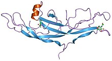
Inhibitor cystine knot (ICK) is a structural motif with a triple stranded antiparallel beta sheet linked by three disulfide bonds, forming a knotted core. The ICK motif can be found under the category of phylum, such as animals and plants. It is often found in many venom peptides such as those of snails, spiders, and scorpions. Peptide K-PVIIA, which contains an ICK, can undergo a successful enzymatic backbone cyclization. The disulfide connectivity and the common sequence pattern of the ICK motif provides the stability of the peptides that support cyclization. [11]
^Wu H, Lustbader JW, Liu Y, Canfield RE, Hendrickson WA (June 1994). "Structure of human chorionic gonadotropin at 2.6 A resolution from MAD analysis of the selenomethionyl protein". Structure. 2 (6): 545–58. doi:10.1016/s0969-2126(00)00054-x. PMID 7922031.
^"Cystine Knots". The Cyclotide Webpage. Archived from the original on 2015-02-05. Retrieved 2019-04-24.
^Sherbet, G.V. (2011), "Growth Factor Families", Growth Factors and Their Receptors in Cell Differentiation, Cancer and Cancer Therapy, Elsevier, pp. 3–5, doi:10.1016/b978-0-12-387819-9.00002-5, ISBN 9780123878199, retrieved 2019-05-01
^Vitt, Ursula A.; Hsu, Sheau Y.; Hsueh, Aaron J. W. (2001-05-01). "Evolution and Classification of Cystine Knot-Containing Hormones and Related Extracellular Signaling Molecules". Molecular Endocrinology. 15 (5): 681–694. doi:10.1210/mend.15.5.0639. ISSN 0888-8809. PMID 11328851.
^Daly NL, Craik DJ (June 2011). "Bioactive cystine knot proteins". Current Opinion in Chemical Biology. 15 (3): 362–8. doi:10.1016/j.cbpa.2011.02.008. PMID 21362584.
^PDB: 1bet; McDonald NQ, Lapatto R, Murray-Rust J, Gunning J, Wlodawer A, Blundell TL (December 1991). "New protein fold revealed by a 2.3-A resolution crystal structure of nerve growth factor". Nature. 354 (6352): 411–4. Bibcode:1991Natur.354..411M. doi:10.1038/354411a0. PMID 1956407. S2CID 4346788.
^Sun PD, Davies DR (1995). "The cystine-knot growth-factor superfamily". Annual Review of Biophysics and Biomolecular Structure. 24 (1): 269–91. doi:10.1146/annurev.bb.24.060195.001413. PMID 7663117.
^Jiang X, Dias JA, He X (January 2014). "Structural biology of glycoprotein hormones and their receptors: insights to signaling". Molecular and Cellular Endocrinology. 382 (1): 424–451. doi:10.1016/j.mce.2013.08.021. PMID 24001578.
^ abIyer S, Acharya KR (November 2011). "Tying the knot: the cystine signature and molecular-recognition processes of the vascular endothelial growth factor family of angiogenic cytokines". The FEBS Journal. 278 (22): 4304–22. doi:10.1111/j.1742-4658.2011.08350.x. PMC 3328748. PMID 21917115.
^Craik DJ, Daly NL, Bond T, Waine C (December 1999). "Plant cyclotides: A unique family of cyclic and knotted proteins that defines the cyclic cystine knot structural motif". Journal of Molecular Biology. 294 (5): 1327–36. doi:10.1006/jmbi.1999.3383. PMID 10600388.
^Kwon, Soohyun; Bosmans, Frank; Kaas, Quentin; Cheneval, Oliver; Cinibear, Anne C; Rosengren, K Johan; Wang, Conan K; Schroeder, Christina I; Craik, David J (19 April 2016). "Efficient enzymatic cyclization of an inhibitory cystine knot‐containing peptide". Biotechnology and Bioengineering. 113 (10): 2202–2212. doi:10.1002/bit.25993. PMC 5526200. PMID 27093300.
A cystineknot is a protein structural motif containing three disulfide bridges (formed from pairs of cysteine residues). The sections of polypeptide that...
folds in the cystineknot motif; the other closely related knots are the growth factor cystineknot (GFCK) and the cyclic cystineknot (CCK; cyclotide)...
Ichthyophthirius multifiliis, a single-celled parasite. Also known as Ich Inhibitor cystineknot Institute of Christ the King Sovereign Priest Intercity Kort, A Dutch...
disulfide bonds. These combined features have been termed the cyclic cystineknot (CCK) motif. To date, over 100 cyclotides have been isolated and characterized...
bridges are the cystineknots, for which two disulfide bridges form a closed, covalent loop, which is threaded by third chain. The term "knot" in the name...
Interleukin 17 family (IL17 family) is a family of pro-inflammatory cystineknot cytokines. They are produced by a group of T helper cell known as T helper...
source of psalmotoxin and vanillotoxin which are classified as inhibitor cystineknot proteins. Psalmotoxin may be of therapeutic use in patients with a stroke...
almost always leads to blindness. It is caused by mutations in the Norrin cystineknot growth factor gene, also referred to as Norrie Disease Pseudoglioma (NDP)...
sub-family of growth factors, the platelet-derived growth factor family of cystine-knot growth factors. They are important signaling proteins involved in both...
substructure have been found in naturally occurring peptides, including: cystineknot peptides, cyclotides or lasso-peptides such as microcin J25. The earliest...
channels. The structure of atracotoxin comprises a core beta region with a cystineknot motif, a feature seen in other neurotoxic polypeptides. Since 1927, records...
protein show six beads of (C and C-like) domains under cryo-EM. the "cystineknot" domain (at the C-terminal end of the protein), which VWF shares with...
tests. Moreover, their complex structure – resembling the inhibitor cystineknot – made them highly stable, explaining how the sting lasts for such a...
gene-1 (USAG-1), an N-glycosylated, secreted protein with a C-terminal, cystine, knot-like domain. This protein functions as a bone morphogenetic protein...
neurons. The structure of versutoxin contains a central beta region with a cystineknot motif, commonly found in other neurotoxic polypeptides, but not found...
contain a cysteine-stabilised triple-stranded beta-sheet with an inhibitor cystineknot motif and show features common to membrane-interactive peptides. Tachystatin...
Pink Toe spider, Avicularia juruensis, which contains the inhibitory cystineknot motif". Frontiers in Microbiology. 3: 324. doi:10.3389/fmicb.2012.00324...
with other tarantula toxins suggests that TLTx also forms and inhibitor cystineknot (ICK) motif. The main difference between the three TLTx subtypes is their...
dysplasia sclerosteosis results from loss of the SOST gene product, a novel cystineknot-containing protein". American Journal of Human Genetics. 68 (3): 577–89...
the VaTxs bind. Vanillotoxins have close homology to other inhibitor cystineknot (ICK) toxins. ICK toxins are best known as blockers of cation channels...
the assassin bug Peirates turpis. The toxin belongs to the inhibitory cystineknot structural family (ICK) that has a core of disulfide bonds with four...
which affects cell proliferation. The structure reveals an inhibitor cystineknot (knottin)-like fold, comprising three beta strands. Jouvensal, L.; Quillien...
is a protein that in humans is encoded by the GPHB5 gene. GPHB5 is a cystineknot-forming polypeptide and a subunit of the dimeric glycoprotein hormone...
have a knottin or inhibitor cystineknot scaffold. The knottin scaffold is a very special disulfide-through-disulfide knot, in which the III-VI disulfide...
"Design and synthesis of heterotrimeric collagen peptides with a built-in cystine-knot. Models for collagen catabolism by matrix-metalloproteases". FEBS Letters...
 Global Information