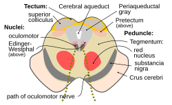
Transverse section of mid-brain at level of superior colliculi. ("Tegmentum" visible center right.)
Section through superior colliculus showing path of oculomotor nerve. (Tegmentum not labeled, but surrounding structures more clearly defined.)
Details
Part of
Midbrain
Identifiers
Latin
Tegmentum Mesencephali
MeSH
D013681
NeuroNames
491
NeuroLex ID
birnlex_1200
Anatomical terms of neuroanatomy
[edit on Wikidata]
The midbrain is anatomically delineated into the tectum (roof) and the tegmentum (floor). The midbrain tegmentum extends from the substantia nigra to the cerebral aqueduct in a horizontal section of the midbrain. It forms the floor of the midbrain that surrounds below the cerebral aqueduct as well as the floor of the fourth ventricle while the midbrain tectum forms the roof of the fourth ventricle. The tegmentum contains a collection of tracts and nuclei with movement-related, species-specific, and pain-perception functions. The general structures of midbrain tegmentum include red nucleus and the periaqueductal grey matter.
Nigrostriatal Pathway and Mesolimbic Pathway in the Dopaminergic System
Unlike the midbrain tectum (which is a sensory structure located posteriorly), the midbrain tegmentum, which locates anteriorly, is related to a number of motor functions. Within the tegmentum, the red nucleus is in charge of motor coordination (specifically for limb movements) and the periaqueductal gray matter (PAG) contains critical circuits for modulating behavioral responses to pains. The substantia nigra (black substance) serves an important role in rewarding behaviors such as approaching desired objects. In addition, the substantia nigra forms reciprocal connections with the basal ganglia which are highly correlated with motor functions and learning.
The midbrain tegmentum is also an important part of the dopaminergic system which is essential for feelings of reward and pleasure. Two regions in the midbrain tegmentum are of particular interest. The first one is the substantia nigra which is an important part of the nigrostriatal pathway. This pathway serves to coordinate motor movements and when left unbalanced, motor deficits would follow. For instance, when the dopamine neurons are lost from the substantia nigra, the condition of extreme muscle rigidity occurs as in the Parkinson's disease. The second region is the ventral tegmental area (VTA; or simply ventral tegmentum) which is at the hub of the mesolimbic pathway. Specifically, the VTA is the origin of dopaminergic cell bodies from which signals reach the anterior parts of the brain (e.g., frontal lobes) as well as the posterior parts (e.g., cerebellum). Because of this pathway regulates the experience of reward and pleasure, it is not surprising to see that food and drugs affect it the most in terms of a loss of impulse control. That is, the mesolimbic pathway is essential in regulating drug addiction. The potential mechanism is through the associative learning of the environmental cues and reward. For instance, through each drug use, individuals increasingly associate the cues related to each drug use (e.g., the room in which the drug is taken or the people with which individuals take drug). Over time, the drug enhances the dopamine-related, classically-conditioned cues associated with drug taking. As a result, later encounters with these cues will produce and heighten dopamine activity and subsequently prompt individuals to crave drugs. Moreover, excessive mesolimbic dopamine activity plays a role in schizophrenia, a behavioral disorder characterized by delusions, hallucinations, blunted emotion, agitation, etc. On the other hand, a lack of mesolimbic dopamine activity may induce deficits in attention.[1]
^Kolb, Bryan; Ian Q. Whishaw. (2015). Fundamentals of Human Neuropsychology (7. ed.). New York, NY: Macmillan. p. 71-G-30. ISBN 978-1-4292-8295-6.{{cite book}}: CS1 maint: multiple names: authors list (link)
and 30 Related for: Midbrain tegmentum information
The midbrain is anatomically delineated into the tectum (roof) and the tegmentum (floor). The midbraintegmentum extends from the substantia nigra to the...
The tegmentum (from Latin for "covering") is a general area within the brainstem. The tegmentum is the ventral part of the midbrain and the tectum is...
principal regions of the midbrain are the tectum, the cerebral aqueduct, tegmentum, and the cerebral peduncles. Rostrally the midbrain adjoins the diencephalon...
is a nucleus situated in the brainstem, spanning the midbraintegmentum and the pontine tegmentum. Its location is one-third of the way from the pedunculopontine...
The pontine tegmentum, or dorsal pons, is located within the brainstem, and is one of two parts of the pons, the other being the ventral pons or basilar...
nucleus (IPN) is an unpaired, ovoid cell group at the base of the midbraintegmentum. It is located in the mesencephalon below the interpeduncular fossa...
tegmental area (VTA) (tegmentum is Latin for covering), also known as the ventral tegmental area of Tsai, or simply ventral tegmentum, is a group of neurons...
from the reticular formation of the lower brain stem through the midbraintegmentum, subthalamus and hypothalamus to the internal capsule." The latter...
"neurofibrillary tangles and neuronal loss in the globus pallidus, hypothalamus, midbraintegmentum, periaqueductal gray matter, striatum, and the substantia nigra"....
sleepiness. This was further narrowed down to show that the central midbraintegmentum is the region that plays a role in cortical activation. Thus, sleep...
anti-cancer drug Ventral tegmental area (in neuroanatomy), part of the midbraintegmentum This disambiguation page lists articles associated with the title...
to press a bar to obtain an injection of opiates directly into the midbraintegmentum or the nucleus accumbens. The same animals do not work to obtain the...
either side of the midbrain and are the frontmost part of the midbrain, and act as the connectors between the rest of the midbrain and the thalamic nuclei...
in the midbrain of healthy, adult humans.[citation needed] Group A10 is the largest group of dopaminergic cells in the ventral midbraintegmentum of rodents...
of the brainstem that in humans and other mammals, lies inferior to the midbrain, superior to the medulla oblongata and anterior to the cerebellum. The...
below the foramen magnum inferiorly. The midbrain is further subdivided into three parts: tectum, tegmentum, and the ventral tegmental area. The tectum...
gray layer of the superior colliculus and more laterally near the midbraintegmentum. Riley HA (1943). An Atlas Of The Basal Ganglia, Brain Stem And Spinal...
forms: hemoglobin and ferritin. The structure is located in the tegmentum of the midbrain next to the substantia nigra and comprises caudal magnocellular...
lesion (infarction, hemorrhage, tumor, or tuberculosis) in the tegmentum of the midbrain and cerebellum. Specifically, the median zone is impaired. It...
of projections from the subicular complex, prefrontal cortex, and midbraintegmentum". J Comp Neurol. 286 (3): 311–36. doi:10.1002/cne.902860303. PMID 2504784...
performed to look for structural lesions in areas such as the thalamus, midbraintegmentum, and substantia nigra. Treatment of a Holmes tremor can fail or is...
a lesion involving the red nucleus and corticospinal tract in the midbraintegmentum. Benedikt is remembered today for his controversial research in criminal...
eye movements. At the level of the caudal midbrain, corticomesencephalic fibers descend through the tegmentum in the medial lemniscus toward the oculomotor...
system. Neurons of the midbraintegmentum of the CF-FM bat brain have been implicated in the DSC mechanism. Neurons of the tegmentum have firing properties...
side of the midbrain, and descends in the lateral part of the brainstem tegmentum. In the spinal cord, it travels through the lateral funiculus of the spinal...
the gray matter located around the cerebral aqueduct within the tegmentum of the midbrain. It projects to the nucleus raphe magnus, and also contains descending...
dorsolateral quadrant of the lateral funiculus, in the lateral tegmentum of the medulla, pons and midbrain. The first-order neuron of the hypothalamospinal tract...
 Global Information