Depression in the temporal bone that articulates with the mandible
Not to be confused with glenoid fossa.
Mandibular fossa
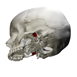
Left temporal bone. Outer surface. (Mandibular fossa labeled at left, third from the top.)
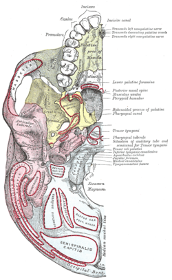
Base of skull. Inferior surface. (Mandibular fossa labeled at center left. Temporal bone is pink.)
Details
Part of
Temporal bone
System
Skeletal
Identifiers
Latin
fossa mandibularis
TA98
A02.1.06.071
TA2
712
FMA
75313
Anatomical terms of bone
[edit on Wikidata]
The mandibular fossa, also known as the glenoid fossa in some dental literature, is the depression in the temporal bone that articulates with the mandible.
The mandibularfossa, also known as the glenoid fossa in some dental literature, is the depression in the temporal bone that articulates with the mandible...
fibrocartilagenous tissue that is positioned between the head of the mandibular condyle and the mandibularfossa of the temporal bone. The temporomandibular joints are...
retromandibular vein, and nerves such as the mandibular nerve (CN V3) and its branches. The boundaries of the infratemporal fossa occur: anteriorly, by the infratemporal...
depression, forming part of the mandibularfossa, for the reception of the condyle of the mandible. The mandibularfossa (glenoid fossa) is bounded, in front,...
the mandibular nerve (also known as the nervus spinosus) is a sensory branch of the mandibular nerve (CN V3) that enters the middle cranial fossa through...
the mandibular jaw position in which the head of the condyle is situated as far anterior and superior as it possibly can within the mandibularfossa/glenoid...
cranium's mandibularfossa. Its upper surface is concavo-convex from before backward, to accommodate itself to the form of the mandibularfossa and the...
tubercle forms the front boundary of the mandibularfossa, and in the fresh state is covered with cartilage. The mandibular condyle normally moves over the articular...
runs from the temporomandibular joint to the tympanic cavity. The mandibularfossa is bounded, in front, by the articular tubercle; behind, by the tympanic...
The middle cranial fossa is formed by the sphenoid bones, and the temporal bones. It lodges the temporal lobes, and the pituitary gland. It is deeper...
through the roof of the glenoid fossa and into the middle cranial fossa is rare. Other rare complications of mandibular trauma include internal carotid...
the mandible is marked in the midline by a faint ridge, indicating the mandibular symphysis, the line of junction of the two halves of the mandible. This...
line Sublingual fossa Submandibular fossa Alveolar part Dental alveoli Ramus of mandible Angle of mandible Mandibular foramen Mandibular canal Mylohyoid...
shortened rostrum, absent alisphenoid canals, and a relatively flat mandibularfossa. Kinkajous have unique morphological characteristics due to their arboreally...
the right permanent mandibular central incisor is known as "41", and the left one is known as "31". The central incisors have fossa on their lingual surfaces...
lateral pterygoid muscle is to pull the head of the condyle out of the mandibularfossa along the articular eminence to protrude the mandible. A concerted...
central fossa from which multiple irregular fissures originate. Their roots are commonly fused together and can be irregular in shape. Mandibular (lower)...
closed about the fifth year, but may persist throughout life. The mandibularfossa is at first extremely shallow, and looks lateral and inferior; it deepens...
arises by two roots: an anterior, directed inward in front of the mandibularfossa, where it expands to form the articular tubercle. a posterior, which...
immediately below the foramen ovale in the infratemporal fossa and on the medial surface of the mandibular nerve. It is functionally associated with the glossopharyngeal...
muscle, to the pterygopalatine fossa. It supplies the deep structures of the face, and may be divided into mandibular, pterygoid, and pterygopalatine...
suggested that the true mandibularfossa was part of the area Lamberton identified as such, at the side of the braincase. The fossa is small and low, suggesting...
through the infratemporal fossa as part of a neurovascular bundle with the inferior alveolar nerve and vein to the mandibular foramen where it enters and...
A trifid mandibular canal variation has also been described. Mandibular nerve and bone. Deep dissection. Anterior view. Infratemporal fossa. Lingual and...
muscle) is a thick, quadrilateral muscle of the face. It is supplied by the mandibular branch of the trigeminal nerve (V). It is important in mastication (chewing)...
the skull). The most important feature is pain, followed by restricted mandibular movement, and noises from the temporomandibular joints (TMJ) during jaw...
notch of temporal bone, and its anterior belly is attached to the digastric fossa of mandible; the two bellies are united by an intermediate tendon which...
 Global Information