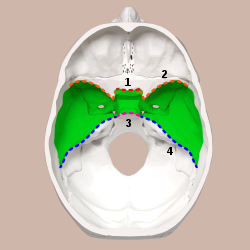
Superior view of the skull base. Middle cranial fossa shown in green.
1: Sphenoidal limbus (anterior margin of the chiasmatic groove)
2: Posterior borders of the lesser wings of the sphenoid
3: Dorsum sellae of the sphenoid bone
4: Superior borders of the petrous part of the temporal bone
Base of the skull. Upper surface. (Middle cranial fossa is the centermost of the three indentations, in pink and yellow.)
Details
Identifiers
Latin
fossa cranii media
MeSH
D035301
TA98
A02.1.00.049
TA2
452
FMA
54369
Anatomical terminology
[edit on Wikidata]
The middle cranial fossa is formed by the sphenoid bones, and the temporal bones. It lodges the temporal lobes, and the pituitary gland.[1][2] It is deeper than the anterior cranial fossa, is narrow medially and widens laterally to the sides of the skull. It is separated from the posterior cranial fossa by the clivus and the petrous crest.
It is bounded in front by the posterior margins of the lesser wings of the sphenoid bone, the anterior clinoid processes, and the ridge forming the anterior margin of the chiasmatic groove; behind, by the superior angles of the petrous portions of the temporal bones and the dorsum sellae; laterally by the temporal squamae, sphenoidal angles of the parietals, and greater wings of the sphenoid. It is traversed by the squamosal, sphenoparietal, sphenosquamosal, and sphenopetrosal sutures.
^Standring, Susan (2020). Gray's Anatomy: The Anatomical Basis of Clinical Practice (42nd ed.). [New York]. ISBN 978-0-7020-7707-4. OCLC 1201341621.{{cite book}}: CS1 maint: location missing publisher (link)
^Mancall, Elliott L.; Brock, David G., eds. (2011). "Cranial Fossae". Gray's Clinical Anatomy. Elsevier Health Sciences. p. 154. ISBN 9781437735802.
and 28 Related for: Middle cranial fossa information
The middlecranialfossa is formed by the sphenoid bones, and the temporal bones. It lodges the temporal lobes, and the pituitary gland. It is deeper than...
A cranialfossa is formed by the floor of the cranial cavity. There are three distinct cranial fossae: Anterior cranialfossa (fossa cranii anterior),...
communicates with the nasal and oral cavities, infratemporal fossa, orbit, pharynx, and middlecranialfossa through eight foramina. It has the following boundaries:...
The posterior cranialfossa is the part of the cranial cavity located between the foramen magnum, and tentorium cerebelli. It is formed by the sphenoid...
The anterior cranialfossa is a depression in the floor of the cranial base which houses the projecting frontal lobes of the brain. It is formed by the...
spread into the infratemporal fossa. This can be surgically removed through the middlecranialfossa. The infratemporal fossa can also be used to approach...
into the middlecranialfossa of the cranial cavity, then exits the cranial cavity through its own canaliculus to reach the infratemporal fossa. Cell bodies...
from the neck into (the middlecranialfossa of) the cranial cavity. The carotid canal is located within the middlecranialfossa, at the petrous part of...
hypophyseal fossa. The sella turcica is located in the sphenoid bone behind the chiasmatic groove and the tuberculum sellae. It belongs to the middlecranial fossa...
to reach the pterygopalatine fossa and form the pterygopalatine ganglion).: 498 During surgery of the middlecranialfossa, manipulation of the dura mater...
through the foramen ovale to enter the cranial cavity and supply the dura mater of the floor of the middlecranialfossa and of the trigeminal cave, and to...
in the sphenoid bone of the skull. It connects the middlecranialfossa and the pterygopalatine fossa. It allows for the passage of the maxillary nerve...
body. The human skull has numerous openings (foramina), through which cranial nerves, arteries, veins, and other structures pass. These foramina vary...
foramen lacerum. The anterior surface forms the posterior part of the middlecranialfossa of the base of the skull, and is continuous with the inner surface...
is a continuation of the tegmen tympani and separates it from the middlecranialfossa. The lateral wall of the antrum is formed by a plate of bone which...
suture) is the cranial suture between the sphenoid bone and the petrous portion of the temporal bone. It is in the middlecranialfossa. This article incorporates...
supply, in the middlecranialfossa the middle meningeal artery and some accessory arteries are responsible for blood supply, the middle meningeal artery...
V3) that enters the middlecranialfossa through either the foramen spinosum or foramen ovale to innervate the meninges of this fossa as well as the mastoid...
bone. Basilar skull fractures are divided into anterior fossa, middlefossa and posterior fossa fractures. Facial fractures often also occur. Diagnosis...
and middle ear cavities are exteriorized so as not to give the chance for the infection or the cholesteatoma to spread into the middlecranialfossa. Since...
Diploe Cranial base Internal surface of cranial base Petrosphenoidal fissure Petro-occipital fissure Anterior cranialfossaMiddlecranialfossa Posterior...
forms the floor of the middlecranialfossa: 508-509 ) at the anterior boundary of the sella turcica (hypophyseal (pituitary) fossa): 509 and posterior...
: 509 It connects the middlecranialfossa (superiorly), and infratemporal fossa (inferiorly). The foramen transmits the middle meningeal artery and vein...
end.[citation needed] Anatomically, it relates superiorly to the middlecranialfossa, posteriorly to the ossicles and facial nerve, inferiorly to the...
 Global Information