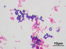
Micrograph of a gram-positive coccus and a gram-negative rod.A Gram stain of mixed Staphylococcus aureus (S. aureus ATCC 25923, gram-positive cocci, in purple) and Escherichia coli (E. coli ATCC 11775, gram-negative bacilli, in red), the most common Gram stain reference bacteria
Gram stain (Gram staining or Gram's method), is a method of staining used to classify bacterial species into two large groups: gram-positive bacteria and gram-negative bacteria. It may also be used to diagnose a fungal infection.[1] The name comes from the Danish bacteriologist Hans Christian Gram, who developed the technique in 1884.[2]
Gram staining differentiates bacteria by the chemical and physical properties of their cell walls. Gram-positive cells have a thick layer of peptidoglycan in the cell wall that retains the primary stain, crystal violet. Gram-negative cells have a thinner peptidoglycan layer that allows the crystal violet to wash out on addition of ethanol. They are stained pink or red by the counterstain,[3] commonly safranin or fuchsine. Lugol's iodine solution is always added after addition of crystal violet to strengthen the bonds of the stain with the cell membrane.
Gram staining is almost always the first step in the identification of a bacterial group. While Gram staining is a valuable diagnostic tool in both clinical and research settings, not all bacteria can be definitively classified by this technique. This gives rise to gram-variable and gram-indeterminate groups.
^"Gram Stain: MedlinePlus Medical Test". medlineplus.gov.
^Colco, R. (2005). "Gram Staining". Current Protocols in Microbiology. Appendix 3 (1): Appendix 3C. doi:10.1002/9780471729259.mca03cs00. ISBN 978-0471729259. PMID 18770544. S2CID 32452815.
^Cite error: The named reference Beveridge_and_Davies_1983 was invoked but never defined (see the help page).
counterstain is stain that makes cells or structures more visible, when not completely visible with the principal stain. Crystal violet stains both Gram positive...
Christian Joachim Gram (13 September 1853 – 14 November 1938) was a Danish bacteriologist noted for his development of the Gramstain, still a standard...
differential staining is the Gramstain. Gramstaining uses two dyes: Crystal violet and Fuchsin or Safranin (the counterstain) to differentiate between Gram-positive...
mycolic acid, in their cell walls which allow them to be stained with Acid-Fast better than a Gram-Stain. The unique ability of mycobacteria to resist decolorization...
to allow the organisms to multiply. If microbial growth is detected, a Gramstain is conducted from the culture bottle to confirm that organisms are present...
inside the vagina should be obtained. These swabs can be tested for: Gramstain which shows the depletion of lactobacilli and overgrowth of Gardnerella...
classify bacteria into Gram-positive bacteria and Gram-negative bacteria. The names originate from the reaction of cells to the Gramstain, a long-standing...
Most pathogenic bacteria can be grown in cultures and identified by Gramstain and other methods. Bacteria grown in this way are often tested to find...
Giemsa stain (/ˈɡiːmzə/), named after German chemist and bacteriologist Gustav Giemsa, is a nucleic acid stain used in cytogenetics and for the histopathological...
characteristic of Gram-positive bacteria. Despite this evolutionary divergence, instances have been reported where U. urealyticum, upon gramstaining, exhibited...
the cell wall in Gram-negative bacteria. The peptidoglycan layer takes up the crystal violet dye and stains purple in the Gramstain. Bacteria within...
The Nugent Score is a Gramstain scoring system for vaginal swabs to diagnose bacterial vaginosis (BV). The Nugent score is calculated by assessing for...
categories: a Gram-positive type which stains purple during Gramstaining and a Gram-negative type which stains pink during Gramstaining. Either type...
management, and safety are unknown. If Gram-negative, oxidase-positive diplococci are visualized on direct Gramstain of urethral pus (male genital infection)...
of ampicillin is recommended to cover Listeria monocytogenes. Once the Gramstain results become available, and the broad type of bacterial cause is known...
fuchsine stain in the Gimenez staining technique and the eosin counterstain to haematoxylin in the H&E stain. In Gramstaining, crystal violet stains only...
conventionally divided into two main groups—gram-positive and gram-negative, based upon their Gram-stain retention property—this classification system...
(haematoxylin-eosin stain, zoom 100×): the large white areas between the muscle fibers are due to gas formation. Gramstain of a muscle biopsy showing Gram-positive...
Gram-positive staining common to class Bacilli. For example, E. coli is a rod-shaped bacterium that can be described as "a bacillus", but it stains Gram-negative...
stain using normal techniques such as simple staining and gramstaining. Special techniques for endospore staining include the Schaeffer–Fulton stain...
absent cell wall structures and do not take up Gramstain in the same manner as gram-negative and gram-positive organisms. Pneumonia Fever Rigors Cough...
bacteria that do not get colored by gram-staining but rather remain colorless: they are neither Gram-positive nor Gram-negative. These include the Chlamydiaceae...
 Global Information