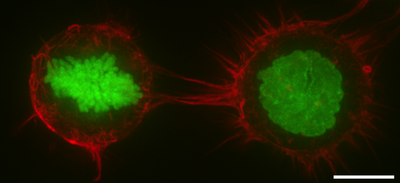
F-actin distribution in the cell cortex as shown by rhodamine phalloidin staining of HeLa cells that constitutively express Histone H2B-GFP to mark chromosomes. F-actin is thus red, while Histone H2B is displayed in green. The left-hand cell is in mitosis, as demonstrated by chromosome condensation, while the right-hand cell is in interphase (as determined by intact cell nucleus) in a suspended state. In both cases, F-actin is enriched around the cell periphery. Scale bar: 10 micrometers.
The cell cortex, also known as the actin cortex, cortical cytoskeleton or actomyosin cortex, is a specialized layer of cytoplasmic proteins on the inner face of the cell membrane. It functions as a modulator of membrane behavior and cell surface properties.[1][2][3] In most eukaryotic cells lacking a cell wall, the cortex is an actin-rich network consisting of F-actin filaments, myosin motors, and actin-binding proteins.[4][5] The actomyosin cortex is attached to the cell membrane via membrane-anchoring proteins called ERM proteins that plays a central role in cell shape control.[1][6] The protein constituents of the cortex undergo rapid turnover, making the cortex both mechanically rigid and highly plastic, two properties essential to its function. In most cases, the cortex is in the range of 100 to 1000 nanometers thick.
In some animal cells, the protein spectrin may be present in the cortex. Spectrin helps to create a network by cross-linked actin filaments.[3] The proportions of spectrin and actin vary with cell type.[7] Spectrin proteins and actin microfilaments are attached to transmembrane proteins by attachment proteins between them and the transmembrane proteins. The cell cortex is attached to the inner cytosolic face of the plasma membrane in cells where the spectrin proteins and actin microfilaments form a mesh-like structure that is continuously remodeled by polymerization, depolymerization and branching.
Many proteins are involved in the cortex regulation and dynamics, including formins, with roles in actin polymerization, Arp2/3 complexes that give rise to actin branching and capping proteins. Due to the branching process and the density of the actin cortex, the cortical cytoskeleton can comprise a highly complex meshwork such as a fractal structure.[8] Specialized cells are usually characterized by a very specific cortical actin cytoskeleton. For example, in red blood cells, the cell cortex consists of a two-dimensional cross-linked elastic network with pentagonal or hexagonal symmetry, tethered to the plasma membrane and formed primarily by spectrin, actin and ankyrin.[9] In neuronal axons, the actin or spectric cytoskeleton forms an array of periodic rings [10] and in the sperm flagellum it forms a helical structure.[11]
In plant cells, the cell cortex is reinforced by cortical microtubules underlying the plasma membrane. The direction of these cortical microtubules determines which way the cell elongates when it grows.
^ abSalbreux G, Charras G, Paluch E (October 2012). "Actin cortex mechanics and cellular morphogenesis". Trends in Cell Biology. 22 (10): 536–45. doi:10.1016/j.tcb.2012.07.001. PMID 22871642.
^ abAlberts, Bruce; Johnson, Alexander; Lewis, Julian; Raff, Martin; Roberts, Keith; Walter, Peter (2002). "Cross-linking Proteins with Distinct Properties Organize Different Assemblies of Actin Filaments". Molecular Biology of the Cell (4th ed.). New York: Garland Science. ISBN 0-8153-3218-1.
^Gunning PW, Ghoshdastider U, Whitaker S, Popp D, Robinson RC (June 2015). "The evolution of compositionally and functionally distinct actin filaments". Journal of Cell Science. 128 (11): 2009–19. doi:10.1242/jcs.165563. PMID 25788699.
^Clark AG, Wartlick O, Salbreux G, Paluch EK (May 2014). "Stresses at the cell surface during animal cell morphogenesis". Current Biology. 24 (10): R484-94. Bibcode:2014CBio...24.R484C. doi:10.1016/j.cub.2014.03.059. PMID 24845681.
^Fehon RG, McClatchey AI, Bretscher A (April 2010). "Organizing the cell cortex: the role of ERM proteins". Nature Reviews. Molecular Cell Biology. 11 (4): 276–87. doi:10.1038/nrm2866. PMC 2871950. PMID 20308985.
^Machnicka B, Grochowalska R, Bogusławska DM, Sikorski AF, Lecomte MC (January 2012). "Spectrin-based skeleton as an actor in cell signaling". Cellular and Molecular Life Sciences. 69 (2): 191–201. doi:10.1007/s00018-011-0804-5. PMC 3249148. PMID 21877118.
^Sadegh S, Higgins JL, Mannion PC, Tamkun MM, Krapf D (2017). "Plasma Membrane is Compartmentalized by a Self-Similar Cortical Actin Meshwork". Physical Review X. 7 (1): 011031. arXiv:1702.03997. Bibcode:2017PhRvX...7a1031S. doi:10.1103/PhysRevX.7.011031. PMC 5500227. PMID 28690919.
^Gov NS (January 2007). "Active elastic network: cytoskeleton of the red blood cell". Physical Review E. 75 (1 Pt 1): 011921. Bibcode:2007PhRvE..75a1921G. doi:10.1103/PhysRevE.75.011921. PMID 17358198.
^Xu K, Zhong G, Zhuang X (January 2013). "Actin, spectrin, and associated proteins form a periodic cytoskeletal structure in axons". Science. 339 (6118): 452–6. Bibcode:2013Sci...339..452X. doi:10.1126/science.1232251. PMC 3815867. PMID 23239625.
^Gervasi MG, Xu X, Carbajal-Gonzalez B, Buffone MG, Visconti PE, Krapf D (June 2018). "The actin cytoskeleton of the mouse sperm flagellum is organized in a helical structure". Journal of Cell Science. 131 (11): jcs215897. doi:10.1242/jcs.215897. PMC 6031324. PMID 29739876.
The cellcortex, also known as the actin cortex, cortical cytoskeleton or actomyosin cortex, is a specialized layer of cytoplasmic proteins on the inner...
Pyramidal cells, or pyramidal neurons, are a type of multipolar neuron found in areas of the brain including the cerebral cortex, the hippocampus, and...
adrenal gland Cellcortex, the region of a cell directly underneath the membrane Cortex (hair), the middle layer of a strand of hair Cortex (botany), the...
lymphocytes, a type of white blood cell, and are primarily made up of B cells and T cells. B cells are mainly found in the outer cortex where they are clustered...
anatomy, the prefrontal cortex (PFC) covers the front part of the frontal lobe of the cerebral cortex. It is the association cortex in the frontal lobe....
function of the reelin positive cells in the layer II of the entorhinal cortex. According to this concept, these cells would be generally organized into...
appearance of the cortex under a microscope. The primary motor cortex contains cells with giant cell bodies known as "Betz cells". These cells were mistakenly...
000. The large, spherical cell bodies of Purkinje cells are packed into a narrow layer (one cell thick) of the cerebellar cortex, called the Purkinje layer...
the neopallium, isocortex, or the six-layered cortex, is a set of layers of the mammalian cerebral cortex involved in higher-order brain functions such...
The visual cortex of the brain is the area of the cerebral cortex that processes visual information. It is located in the occipital lobe. Sensory input...
the primary motor cortex. These neurons are the largest in the central nervous system, sometimes reaching 100 μm in diameter. Betz cells are upper motor...
microtubules from the spindle poles carry a furrow-inducing signal to the cellcortex, where signals from two poles are somehow focused into a ring at the...
The adrenal cortex is the outer region and also the largest part of the adrenal gland. It is divided into three separate zones: zona glomerulosa, zona...
structure Bangstad syndrome CellcortexCell damage, including damage to cell membrane Cell theory Cytoneme Elasticity of cell membranes Gram-positive bacteria...
the diffusion of molecules from one compartment of the cell to another, or in the cellcortex as a barrier to the diffusion of membrane-bound proteins...
Basket cells are inhibitory GABAergic interneurons of the brain, found throughout different regions of the cortex and cerebellum. Basket cells are multipolar...
system; cortical spiny stellate cells are found in layer IVC of the primary visual cortex. In the somatosensory barrel cortex of mice and rats, glutamatergic...
processes such as cell division. The concentrated inner area is called the endoplasm and the outer layer is called the cellcortex, or ectoplasm. Movement...
to cell size asymmetry is spindle-independent. The mechanism instead relies on the spatial and temporal organization of myosin on the cellcortex and...
neurons in the cerebral cortex. RGPs also produce certain lineages of glia, including astrocytes and oligodendrocytes. Their cell bodies (somata) reside...
and myosin (actomyosin) into a contractile homogeneous cellcortex that 1) rigidifies the cell periphery and 2) facilitates generation of intracellular...
of the volume of the cortex. In the zona fasciculata, cells are arranged in columns radially oriented towards the medulla. Cells contain numerous lipid...
movements. Primary motor cortex is defined anatomically as the region of cortex that contains large neurons known as Betz cells, which, along with other...
The ventromedial prefrontal cortex (vmPFC) is a part of the prefrontal cortex in the mammalian brain. The ventral medial prefrontal is located in the...
Basket cells, interneurons that form a dense plexus of terminals around the soma of target cells, found in the cortex and cerebellum Betz cells, large...
adrenal cortex to release glucocorticoids and plays an important role in the stress response. The primary function of the corticotropic cells is to produce...
 Global Information