Fetus of about eight weeks, enclosed in the amnion. (Vitelline duct labeled at lower right.)
Sketches in profile of two stages in the development of the human digestive tube. (Vitelline duct labeled on bottom image.)
Details
Days
28
Precursor
midgut, yolk sac
Identifiers
Latin
ductus vitellinus
MeSH
D014816
Anatomical terminology
[edit on Wikidata]
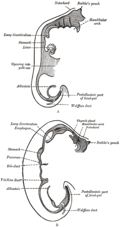
In the human embryo, the vitelline duct, also known as the vitellointestinal duct,[1] the yolk stalk,[1] the omphaloenteric duct,[1] or the omphalomesenteric duct,[1] is a long narrow tube that joins the yolk sac to the midgut lumen of the developing fetus.[2] It appears at the end of the fourth week, when the yolk sac (also known as the umbilical vesicle) presents the appearance of a small pear-shaped vesicle.
^ abcdElsevier, Dorland's Illustrated Medical Dictionary, Elsevier.
^Le, Tao; Bhushan, Vikas; Vasan, Neil (2010). First Aid for the USMLE Step 1: 2010 20th Anniversary Edition. USA: The McGraw-Hill Companies, Inc. pp. 122. ISBN 978-0-07-163340-6.
embryo, the vitellineduct, also known as the vitellointestinal duct, the yolk stalk, the omphaloenteric duct, or the omphalomesenteric duct, is a long...
embryo and its yolk sac Vitelline cyst, a developmental defect relating to the closure of the vitellineductVitellineduct, a tube that joins the yolk...
umbilical vesicle), into the digestive tube by a long narrow tube, the vitellineduct. Rarely, the yolk sac can be seen in the afterbirth as a small, somewhat...
fetus the ileum is connected to the navel by the vitellineduct. In roughly 2−4% of humans, this duct fails to close during the first seven weeks after...
midgut, remains temporally connected to the yolk sac by means of the vitellineduct. The foregut gives rise to the esophagus, the trachea, lung buds, the...
falciform ligament, median umbilical ligament, or a remnant of the vitellineduct. Sister Mary Joseph nodule is associated with multiple peritoneal metastases...
Omphalomesenteric duct cysts (ODC, also known as an omphalomesenteric duct remnant or vitelline cyst) are developmental defects relating to the closure...
detected by ultrasound. All essential organs have at least begun. The vitellineduct normally closes. Gestational age: 9 weeks and 0 days until 11 weeks...
primitive gut. The yolk sac remains connected to the gut tube via the vitellineduct. Usually, this structure regresses during development; in cases where...
involutes before birth. Uncommonly, the yolk sac may persist as the vitellineduct and cause a congenital out pouching of the digestive tract called Meckel's...
branched, tubular ovary. From here, a short oviduct passes to the vitellineduct. This duct connects, via a junction, the ovaries, the uterus, and the yolk...
level of the male accessory gland reservoir to form the ovo-vitellineduct. The ovo-vitellineduct continues along the midline before connecting with the ootype...
of the amnion from the umbilical ring (surrounding the roots of the vitellineduct and connecting stalk) creates a tube with a covering of amniotic membrane...
fertilization occurs. The ootype is connected via a pair of ducts to a number of vitellineducts that produce yolk. After the egg is surrounded by yolk, its...
vitellarium extends from the level of the vitelline reservoir; a pair of vitellineducts form the vitelline reservoir. The vaginal pore is large and guarded...
form the common hepatic duct. The cystic duct from the gallbladder joins with the common hepatic duct to form the common bile duct. The biliary system and...
immediately proximal to vagina and anterior to ootype. Bilateral vitellineducts not observed; vitellarium absent in regions of other reproductive organs...
"Barth's hernia": A hernia of the loops of intestine between a persistent vitellineduct and the serosa of the abdominal wall. Jean Baptiste Philippe Barth @...
and reproduction is stopped. It causes damage of the integument and vitellineduct. Blair DM (1958). "Lucanthone hydrochloride; a review". Bulletin of...
cuticularized margins, and each row is connected with the reticular vitellineducts of its own side, an ovary, and 40-65 large testes pre, para and post-ovarian...
uterus, vitellineduct, ovary, and vitellaria. They also have flame cells that function as a kidney and remove waste material. A short duct that opens...
uterus, vitellineduct, ovary, and vitellaria. They also have flame cells that function as a kidney and remove waste material. A short duct that opens...
but are free in the rest of their extent. It receives blood from the vitelline vein, umbilical vein and common cardinal vein.[citation needed] The sinus...
body midline immediately anterior to ootype. Bilateral and common vitellineducts not observed; vitellarium absent in regions of other reproductive organs...
immediately anterior to ootype and Mehlis' gland. Bilateral and common vitellineducts not observed; vitellarium dense, absent in regions of other reproductive...
branches at the front of the aorta consist of the vitelline arteries and umbilical arteries. The vitelline arteries form the celiac, superior and inferior...
week, the sinus venosus receives blood from the three major veins: the vitelline, the umbilical and the common cardinal veins. During the first two months...
fish-eating mammals, including humans. In humans, it infects the common bile duct and gall bladder, feeding on bile. It was discovered by British physician...
 Global Information