 Global Information
Bilaminar embryonic disc information
| Bilaminar embryonic disc | |
|---|---|
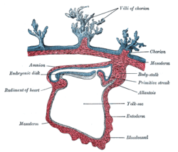
 Section through the embryo. (Bilaminar disc is labeled as embryonic disk.) | |
 Embryonic disc at implantation site with epiblast expanded to form the amniotic cavity | |
| Details | |
| Days | 13 |
| Precursor | Inner cell mass |
| Gives rise to | Human embryo |
| Identifiers | |
| TE | embryonic disc_by_E6.0.1.1.3.0.1 E6.0.1.1.3.0.1 |
| Anatomical terminology [edit on Wikidata] | |
The bilaminar embryonic disc, bilaminar blastoderm or embryonic disc is the distinct two-layered structure of cells formed in an embryo. In the development of the human embryo this takes place by day eight. It is formed when the inner cell mass, also known as the embryoblast, forms a bilaminar disc of two layers, an upper layer called the epiblast (primitive ectoderm) and a lower layer called the hypoblast (primitive endoderm), which will eventually form into fetus.[1][2][3] These two layers of cells are stretched between two fluid-filled cavities at either end: the primitive yolk sac and the amniotic sac.
The epiblast is adjacent to the trophoblast and made of columnar epithelial cells; the hypoblast is closest to the blastocoel (blastocystic cavity) and made of cuboidal cells. As the two layers become evident, a basement membrane forms between the layers. This distinction of layers of the bilaminar disc defines the primitive dorso ventral axis and polarity in embryogenesis.[2]
The epiblast migrates away from the trophoblast downwards, forming the amniotic cavity in between, the lining of which is formed from amnioblasts developed from the epiblast. The hypoblast is pushed down and forms the yolk sac (exocoelomic cavity) lining. Some hypoblast cells migrate along the inner cytotrophoblast lining of the blastocoel, secreting an extracellular matrix along the way. These hypoblast cells and extracellular matrix are called Heuser's membrane (or the exocoelomic membrane), and they cover the blastocoel to form the yolk sac (or exocoelomic cavity). Cells of the hypoblast migrate along the outer edges of this reticulum and form the extraembryonic mesoderm; this disrupts the extraembryonic reticulum. Soon pockets form in the reticulum, which ultimately coalesce to form the chorionic cavity (extraembryonic coelom).
- ^ Sadler, T. W. (2010). Langman's medical embryology (11th ed.). Philadelphia: Lippincott William & Wilkins. p. 54. ISBN 9780781790697.
- ^ a b Schoenwolf, Gary C. (2015). Larsen's human embryology (Fifth ed.). Philadelphia, PA. p. 47. ISBN 9781455706846.
{{cite book}}: CS1 maint: location missing publisher (link) - ^ "27.3B: Bilaminar Embryonic Disc Development". Medicine LibreTexts. 24 July 2018. Retrieved 12 June 2022.