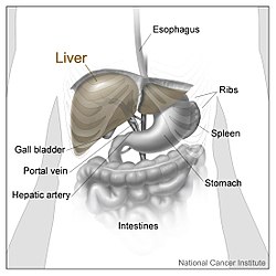
The liver is divided into four lobes. This image shows the large right lobe and a smaller left lobe separated by the falciform ligament.
1: Right lobe of liver 2: Left lobe of liver 3: Quadrate lobe of liver 4: Round ligament of liver 5: Falciform ligament 6: Caudate lobe of liver 7: Inferior vena cava 8: Common bile duct 9: Hepatic artery 10: Portal vein 11: Cystic duct 12: Common hepatic duct 13: Gallbladder
Details
Identifiers
Latin
lobus hepatis
Anatomical terminology
[edit on Wikidata]
In human anatomy, the liver is divided grossly into four parts or lobes: the right lobe, the left lobe, the caudate lobe, and the quadrate lobe. Seen from the front – the diaphragmatic surface – the liver is divided into two lobes: the right lobe and the left lobe. Viewed from the underside – the visceral surface – the other two smaller lobes, the caudate lobe and the quadrate lobe, are also visible.[1] The two smaller lobes, the caudate lobe and the quadrate lobe, are known as superficial or accessory lobes, and both are located on the underside of the right lobe.[2]
The falciform ligament, visible on the front of the liver, makes a superficial division of the right and left lobes of the liver. From the underside, the two additional lobes are located on the right lobe.[2] A line can be imagined running from the left of the vena cava and all the way forward to divide the liver and gallbladder into two halves.[3] This line is called Cantlie's line and is used to mark the division between the two lobes.[4]
Other anatomical landmarks exist, such as the ligamentum venosum and the round ligament of the liver (ligamentum teres), which further divide the left side of the liver in two sections. An important anatomical landmark, the porta hepatis, also known as the transverse fissure of the liver, divides this left portion into four segments, which can be numbered in Roman numerals starting at the caudate lobe as I in an anticlockwise manner. From this parietal view, seven segments can be seen, because the eighth segment is only visible in the visceral view.[5]
Labeled human liver
^Karanjia N. "Anatomy of the Liver". Liver.co.uk. Retrieved 2015-06-26.
^ abMoore KL, Dalley AF, Agur AM (2018). Clinically Oriented Anatomy (Eighth ed.). Philadelphia Baltimore New York London Buenos Aires Hong Kong Sydney Tokyo: Wolters Kluwer. pp. 493–498. ISBN 978-1-4963-5404-4.
^Renz JF, Kinkhabwala M (2014). "Surgical Anatomy of the Liver". In Busuttil RW, Klintmalm GB (eds.). Transplantation of the Liver. Elsevier. pp. 23–39. ISBN 978-1-4557-5383-3.
^Mudgal P, Hacking C, Di Muzio B, et al. "Cantlie's line | Radiology Reference Article". Radiopaedia.org. Retrieved 2015-06-26.
^Kuntz E, Kuntz HD (2009). "Liver resection". Hepatology: Textbook and Atlas (3rd ed.). Springer. pp. 900–903. ISBN 978-3-540-76839-5.
the liver is divided into two lobes: the right lobe and the left lobe. Viewed from the underside – the visceral surface – the other two smaller lobes, the...
division of the liver into a left and right lobe. From below, the two additional lobes are located between the right and left lobes, one in front of the other...
from the caudate lobe. There are four lobesof the liver. The Couinaud classification ofliver anatomy then further divides the liver into eight functionally...
different from the lobes of liver: they are the smaller divisions of the lobes. The two-dimensional microarchitecture of the liver can be viewed from different...
grey-blue organ that sits in a shallow depression below the right lobeof the liver. In adults, the gallbladder measures approximately 7 to 10 centimetres...
fissure of the liver is a short but deep fissure, about 5 cm long, extending transversely beneath the left portion of the right lobeof the liver, nearer...
involvement of both lobesofliver, portal vein invasion and a lower median survival rate. The liver is responsible for the production of the vast majority of coagulation...
ligament of the liver, ligamentum teres or ligamentum teres hepatis is a ligament that forms part of the free edge of the falciform ligament of the liver. It...
attaches the liver to the front body wall and divides the liver into the left lobe and right lobe. The falciform ligament is a broad and thin fold of peritoneum...
tract. It is formed by the union of the right hepatic duct (which drains bile from the right functional lobeof the liver) and the left hepatic duct (which...
system) refers to the liver, gallbladder and bile ducts, and how they work together to make, store and secrete bile. Bile consists of water, electrolytes...
the neck (inferior). Bile is required for the digestion of food and is secreted by the liver into passages that carry bile toward the hepatic duct. It...
Hepatomegaly is enlargement of the liver. It is a non-specific medical sign, having many causes, which can broadly be broken down into infection, hepatic...
arrangement may have the purpose of directing bile flow distally instead of back towards the liver). The inner surface of the cystic duct features spiral...
Medical Center Anatomy image:7957 at the SUNY Downstate Medical Center Liver at The Anatomy Lesson by Wesley Norman (Georgetown University) (biliarysystem)...
the thoracic cavity The bare area of the liver is found on the posterosuperior surface of the right lobeof the liver. This lies close to the thoracic...
majority of pancreatic tissue has a digestive role. The cells with this role form clusters (Latin: acini) around small ducts, and are arranged in lobes that...
Kupffer–Browicz cells, are specialized cells localized in the liver within the lumen of the liver sinusoids and are adhesive to their endothelial cells which...
A hepatocyte is a cell of the main parenchymal tissue of the liver. Hepatocytes make up 80% of the liver's mass. These cells are involved in: Protein...
also known as liver cirrhosis or hepatic cirrhosis, and end-stage liver disease, is the impaired liver function caused by the formation of scar tissue known...
surface of the liver between the caudate and main parts of the left lobe. It is grouped with the liver in Terminologia Anatomica. Ligamentum teres Ligamentum...
The pancreatic duct or duct of Wirsung (also, the major pancreatic duct due to the existence of an accessory pancreatic duct) is a duct joining the pancreas...
digestive system consists of the gastrointestinal tract plus the accessory organs of digestion (the tongue, salivary glands, pancreas, liver, and gallbladder)...
vein, which is then implanted in the liver. There is a risk of portal venous branch thrombosis and the low value of islet survival a few minutes after transplantation...
cells), are pericytes found in the perisinusoidal space of the liver, also known as the space of Disse (a small area between the sinusoids and hepatocytes)...
production of silver stools and can be indicative of cancer of the Ampulla of Vater. The silver-colored stool is a combination of the white stool of obstructive...
A liver sinusoid is a type of capillary known as a sinusoidal capillary, discontinuous capillary or sinusoid, that is similar to a fenestrated capillary...
as part of a collateral circulation that occurs to drain blood from the abdomen as a result of portal hypertension, usually as a result ofliver diseases...
causes it to rotate to the left, and the liver and stomach undergo a lot of growth. This makes the two parts of the pancreas rotate around the duodenum...
 Global Information