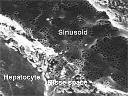
Sinusoid of a rat liver with fenestrated endothelial cells. Fenestrae are approx 100 nm diameter, and the sinusoidal width 5 µm.
Basic liver structure
Details
Drains from
Hepatic portal vein
Drains to
Central veins of liver
Identifiers
Latin
vas sinusoideum
TH
H3.04.05.0.00014
FMA
17543
Anatomical terminology
[edit on Wikidata]
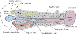
A liver sinusoid is a type of capillary known as a sinusoidal capillary, discontinuous capillary or sinusoid, that is similar to a fenestrated capillary, having discontinuous endothelium that serves as a location for mixing of the oxygen-rich blood from the hepatic artery and the nutrient-rich blood from the portal vein.[1]
The liver sinusoid has a larger caliber than other types of capillaries and has a lining of specialised endothelial cells known as the liver sinusoidal endothelial cells (LSECs), and Kupffer cells.[2] The cells are porous and have a scavenging function.[3] The LSECs make up around half of the non-parenchymal cells in the liver and are flattened and fenestrated.[4] LSECs have many fenestrae that gives easy communication between the sinusoidal lumen and the space of Disse. They play a part in filtration, endocytosis, and in the regulation of blood flow in the sinusoids.[5]
The Kupffer cells can take up and destroy foreign material such as bacteria. Hepatocytes are separated from the sinusoids by the space of Disse. Hepatic stellate cells are present in the space of Disse and are involved in scar formation in response to liver damage.
Defenestration happens when LSECs are lost rendering the sinusoid as an ordinary capillary. This process precedes fibrosis.[6]
^SIU SOM Histology GI
^Brunt, EM; et al. (June 2014). "Pathology of the liver sinusoids". Histopathology. 64 (7): 907–20. doi:10.1111/his.12364. PMID 24393125. S2CID 12709169.
^DeLeve, LD (November 2007). "Hepatic microvasculature in liver injury". Seminars in Liver Disease. 27 (4): 390–400. doi:10.1055/s-2007-991515. PMID 17979075.
^Xing, Y; Zhao, T; Gao, X; Wu, Y (18 February 2016). "Liver X receptor α is essential for the capillarization of liver sinusoidal endothelial cells in liver injury". Scientific Reports. 6: 21309. Bibcode:2016NatSR...621309X. doi:10.1038/srep21309. PMC 4758044. PMID 26887957.
^Arii, S; Imamura, M (2000). "Physiological role of sinusoidal endothelial cells and Kupffer cells and their implication in the pathogenesis of liver injury". Journal of Hepato-biliary-pancreatic Surgery. 7 (1): 40–8. doi:10.1007/s005340050152. PMID 10982590.
^Xie, G; Wang, X; Wang, L; Wang, L; Atkinson, RD; Kanel, GC; Gaarde, WA; Deleve, LD (April 2012). "Role of differentiation of liver sinusoidal endothelial cells in progression and regression of hepatic fibrosis in rats". Gastroenterology. 142 (4): 918–927.e6. doi:10.1053/j.gastro.2011.12.017. PMC 3618963. PMID 22178212.
A liversinusoid is a type of capillary known as a sinusoidal capillary, discontinuous capillary or sinusoid, that is similar to a fenestrated capillary...
capillaries known as liversinusoids, which then lead to hepatic lobules. Hepatic lobules are the functional units of the liver. Each lobule is made up...
between tissue and the capillary blood, and sinusoids, a type of open-pore capillary found in the liver, bone marrow, anterior pituitary gland, and brain...
Liver sinusoidal endothelial cells (LSECs) form the lining of the smallest blood vessels in the liver, also called the hepatic sinusoids. LSECs are highly...
perisinusoidal space (or space of Disse) is a location in the liver between a hepatocyte and a sinusoid. It contains the blood plasma. Microvilli of hepatocytes...
In human anatomy, the liver is divided grossly into four parts or lobes: the right lobe, the left lobe, the caudate lobe, and the quadrate lobe. Seen...
the liver is the first organ to absorb nutrients just taken in by the intestines. After draining into the liversinusoids, blood from the liver is drained...
the accessory organs of digestion (the tongue, salivary glands, pancreas, liver, and gallbladder). Digestion involves the breakdown of food into smaller...
(one vein at each lobule center). They receive the blood mixed in the liversinusoids and return it to circulation via the hepatic veins. The circulation...
the liver: including the sinusoids, the space of Disse, and other vascular structures, which leads to altered resistance to blood flow in the liver, and...
A liver segment is one of eight segments of the liver as described in the widely used Couinaud classification (named after Claude Couinaud) in the anatomy...
Kupffer–Browicz cells, are specialized cells localized in the liver within the lumen of the liversinusoids and are adhesive to their endothelial cells which make...
(inferior). Bile is required for the digestion of food and is secreted by the liver into passages that carry bile toward the hepatic duct. It joins the cystic...
of the liver, ligamentum teres or ligamentum teres hepatis is a ligament that forms part of the free edge of the falciform ligament of the liver. It connects...
the adult human liver. Binucleate cells are also common. Hepatocytes are organised into plates separated by vascular channels (sinusoids), an arrangement...
fissure of the liver is a short but deep fissure, about 5 cm long, extending transversely beneath the left portion of the right lobe of the liver, nearer its...
found in the perisinusoidal space of the liver, also known as the space of Disse (a small area between the sinusoids and hepatocytes). The stellate cell is...
of liver, or hepatic lobules, are small divisions of the liver defined at the microscopic scale. The hepatic lobule is a building block of the liver tissue...
The biliary tract (also biliary tree or biliary system) refers to the liver, gallbladder and bile ducts, and how they work together to make, store and...
area of the liver (nonperitoneal area) is a large triangular area on the diaphragmatic surface of the liver. It is the only part of the liver with no peritoneal...
Medical Center Anatomy image:7957 at the SUNY Downstate Medical Center Liver at The Anatomy Lesson by Wesley Norman (Georgetown University) (biliarysystem)...
the liver, although the structure and position of the gallbladder can vary significantly among animal species. It receives bile, produced by the liver, via...
cerebrum. Finally, albumin leads the indirect bilirubin to the liver. In the liversinusoid, albumin disassociates with the indirect bilirubin and returns...
growth of the wall of the stomach causes it to rotate to the left, and the liver and stomach undergo a lot of growth. This makes the two parts of the pancreas...
SUNY Downstate Medical Center - "Stomach, Spleen and Liver: The Gallbladder and the Bile System" liver at The Anatomy Lesson by Wesley Norman (Georgetown...
the creation of glucose and the breakdown of glycogen to glucose in the liver. It also decreases the uptake of glucose in fat and muscle. Glucagon release...
right functional lobe of the liver) and the left hepatic duct (which drains bile from the left functional lobe of the liver). The duct is about 3 cm long...
islets are transplanted into a portal vein, which is then implanted in the liver. There is a risk of portal venous branch thrombosis and the low value of...
 Global Information