Global Information Lookup Global Information

Electroencephalography information


Electroencephalography
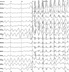
Epileptic spike and wave discharges monitored EEG
[edit on Wikidata]

Electroencephalography (EEG) is a method to record an electrogram of the spontaneous electrical activity of the brain. The biosignals detected by EEG have been shown to represent the postsynaptic potentials of pyramidal neurons in the neocortex and allocortex.[1] It is typically non-invasive, with the EEG electrodes placed along the scalp (commonly called "scalp EEG") using the International 10–20 system, or variations of it. Electrocorticography, involving surgical placement of electrodes, is sometimes called "intracranial EEG". Clinical interpretation of EEG recordings is most often performed by visual inspection of the tracing or quantitative EEG analysis.

Voltage fluctuations measured by the EEG bioamplifier and electrodes allow the evaluation of normal brain activity. As the electrical activity monitored by EEG originates in neurons in the underlying brain tissue, the recordings made by the electrodes on the surface of the scalp vary in accordance with their orientation and distance to the source of the activity. Furthermore, the value recorded is distorted by intermediary tissues and bones, which act in a manner akin to resistors and capacitors in an electrical circuit. This means not all neurons will contribute equally to an EEG signal, with an EEG predominately reflecting the activity of cortical neurons near the electrodes on the scalp. Deep structures within the brain further away from the electrodes will not contribute directly to an EEG; these include the base of the cortical gyrus, mesial walls of the major lobes, hippocampus, thalamus, and brain stem.[2]

A healthy human EEG will show certain patterns of activity that correlate with how awake a person is. The range of frequencies one observes are between 1 and 30 Hz, and amplitudes will vary between 20 and 100 μV. The observed frequencies are subdivided into various groups: alpha (8–13 Hz), beta (13–30 Hz), delta (0.5–4 Hz), and theta (4–7 Hz). Alpha waves are observed when a person is in a state of relaxed wakefulness and are mostly prominent over the parietal and occipital sites. During intense mental activity, beta waves are more prominent in frontal areas as well as other regions. If a relaxed person is told to open their eyes, one observes alpha activity decreasing and an increase in beta activity. Theta and delta waves are not seen in wakefulness, and if they are, it is a sign of brain dysfunction.[2]

EEG can detect abnormal electrical discharges such as sharp waves, spikes, or spike-and-wave complexes that are seen in people with epilepsy; thus, it is often used to inform the medical diagnosis. EEG can detect the onset and spatio-temporal (location and time) evolution of seizures and the presence of status epilepticus. It is also used to help diagnose sleep disorders, depth of anesthesia, coma, encephalopathies, cerebral hypoxia after cardiac arrest, and brain death. EEG used to be a first-line method of diagnosis for tumors, stroke, and other focal brain disorders,[3][4] but this use has decreased with the advent of high-resolution anatomical imaging techniques such as magnetic resonance imaging (MRI) and computed tomography (CT). Despite its limited spatial resolution, EEG continues to be a valuable tool for research and diagnosis. It is one of the few mobile techniques available and offers millisecond-range temporal resolution, which is not possible with CT, PET, or MRI.

Derivatives of the EEG technique include evoked potentials (EP), which involves averaging the EEG activity time-locked to the presentation of a stimulus of some sort (visual, somatosensory, or auditory). Event-related potentials (ERPs) refer to averaged EEG responses that are time-locked to more complex processing of stimuli; this technique is used in cognitive science, cognitive psychology, and psychophysiological research.

  1. ^ Amzica F, Lopes da Silva FH (November 2017). Schomer DL, Lopes da Silva FH (eds.). Cellular Substrates of Brain Rhythms. Vol. 1. Oxford University Press. doi:10.1093/med/9780190228484.003.0002.
  2. ^ a b Eric R. Kandel, John Koester, Sarah Mack, Steven Siegelbaum (2021). Principles of neural science (6th ed.). New York: McGraw-Hill Companies. p. 1450. ISBN 978-1-259-64223-4. OCLC 1199587061.
  3. ^ "EEG: MedlinePlus Medical Encyclopedia". medlineplus.gov. Archived from the original on July 5, 2016. Retrieved July 24, 2022.
  4. ^ Chernecky CC, Berger BJ (2013). Laboratory tests and diagnostic procedures (6th ed.). St. Louis, Mo.: Elsevier. ISBN 978-1-4557-0694-5.

and 25 Related for: Electroencephalography information

Request time (Page generated in 0.5698 seconds.)

Electroencephalography

Last Update:

Electroencephalography (EEG) is a method to record an electrogram of the spontaneous electrical activity of the brain. The biosignals detected by EEG have...

Word Count : 15276

Quantitative electroencephalography

Last Update:

Quantitative electroencephalography (qEEG or QEEG) is a field concerned with the numerical analysis of electroencephalography (EEG) data and associated...

Word Count : 607

Neurology

Last Update:

vessels of the head and neck. Neurophysiologic studies, including electroencephalography (EEG), needle electromyography (EMG), nerve conduction studies (NCSs)...

Word Count : 2504

Electrocorticography

Last Update:

Electrocorticography (ECoG), a type of intracranial electroencephalography (iEEG), is a type of electrophysiological monitoring that uses electrodes placed...

Word Count : 3522

Corpus callosum

Last Update:

Electroencephalography is used to find the source of electrical activity causing a seizure as part of the surgical evaluation for a corpus callosotomy...

Word Count : 3605

Electroconvulsive therapy

Last Update:

Electroconvulsive therapy MECTA spECTrum 5000Q with electroencephalography (EEG) in a modern ECT suite Other names Electroshock therapy ICD-10-PCS GZB...

Word Count : 12279

Fetal EEG

Last Update:

Fetal electroencephalography, also known as prenatal EEG includes any recording of electrical fluctuations arising from the brain of a fetus.  Doctors...

Word Count : 1515

Hans Berger

Last Update:

was a German psychiatrist. He is best known as the inventor of electroencephalography (EEG) in 1924, which is a method used for recording the electrical...

Word Count : 2718

Electrogram

Last Update:

An electrogram (EGM) is a recording of electrical activity of organs such as the brain and heart, measured by monitoring changes in electric potential...

Word Count : 375

Biological basis of personality

Last Update:

Method Function Significance Electroencephalography (EEG) This method measures electrical activity on the surface of the brain through the scalp, and has...

Word Count : 5399

Amplitude integrated electroencephalography

Last Update:

Amplitude integrated electroencephalography (aEEG), cerebral function monitoring (CFM) or continuous electroencephalogram (CEEG) is a technique for monitoring...

Word Count : 358

Seizure

Last Update:

tentatively increases the likelihood that a seizure was the cause. An electroencephalography is only recommended in those who likely had an epileptic seizure...

Word Count : 7089

Hippocampus

Last Update:

"Hippocampal electrical activity and voluntary movement in the rat". Electroencephalography and Clinical Neurophysiology. 26 (4): 407–418. doi:10.1016/0013-4694(69)90092-3...

Word Count : 14840

Attention deficit hyperactivity disorder

Last Update:

rating scale, are used in the screening and evaluation of ADHD. Electroencephalography is not accurate enough to make an ADHD diagnosis. As with many other...

Word Count : 26796

List of neurological research methods

Last Update:

following is a list of neuroimaging methods: Electroencephalography (EEG) Quantitative electroencephalography (QEEG) Stereoelectroencephalography (SEEG)...

Word Count : 99

Boxing

Last Update:

Thompson, Jay S., Kim, Kyung H.: "Boxers: Computed Axial Tomography, Electroencephalography and Neurological Evaluation." Journal of the American Medical Association...

Word Count : 14390

Medical imaging

Last Update:

techniques that are not primarily designed to produce images, such as electroencephalography (EEG), magnetoencephalography (MEG), electrocardiography (ECG),...

Word Count : 7144

Body reactivity

Last Update:

Body reactivity is usually understood as an organism's functional ability of its body to react adequately in response to influence the environment. It...

Word Count : 862

Brain activity and meditation

Last Update:

moment. Electroencephalography (EEG) has been used in many studies as a primary method for evaluating the meditating brain. Electroencephalography uses electrical...

Word Count : 2393

Brain

Last Update:

generate can be large enough to detect outside the skull, using electroencephalography (EEG) or magnetoencephalography (MEG). EEG recordings, along with...

Word Count : 15425

External occipital protuberance

Last Update:

the occipital bone) is used as a landmark in the 10-20 system in electroencephalography (EEG) recording. Extending laterally from it on either side is the...

Word Count : 233

Honda

Last Update:

brainwaves to move ASIMO. The system uses a helmet covered with electroencephalography and near-infrared spectroscopy sensors that monitor electrical brainwaves...

Word Count : 12538

Oneiroid syndrome

Last Update:

oneiroid syndrome, there were crude pathological changes in the electroencephalography (EEG). Clouding of consciousness Delirium Марилов, В. В. (2002)...

Word Count : 2009

Human brain

Last Update:

Medical imaging technologies such as functional neuroimaging, and electroencephalography (EEG) recordings are important in studying the brain. The medical...

Word Count : 18798

Psychology

Last Update:

Single-Channel Recordings to Brain-Mapping Devices: The Impact of Electroencephalography on Experimental Psychology" (PDF). History of Psychology. 8 (1):...

Word Count : 26512

PDF Search Engine © AllGlobal.net