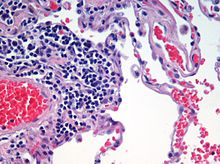
A stained histologic specimen, sandwiched between a glass microscope slide and coverslip, mounted on the stage of a light microscope.Microscopic view of a histologic specimen of human lung tissue stained with hematoxylin and eosin.
Automated tissue image analysis or histopathology image analysis (HIMA) is a process by which computer-controlled automatic test equipment is used to evaluate tissue samples, using computations to derive quantitative measurements from an image to avoid subjective errors.
In a typical application, automated tissue image analysis could be used to measure the aggregate activity of cancer cells in a biopsy of a cancerous tumor taken from a patient. In breast cancer patients, for example, automated tissue image analysis may be used to test for high levels of proteins known to be present in more aggressive forms of breast cancers.
and 25 Related for: Automated tissue image analysis information
Automatedtissueimageanalysis or histopathology imageanalysis (HIMA) is a process by which computer-controlled automatic test equipment is used to evaluate...
evidence that automated systems can perform better than humans. In addition, automated systems are unbiased, unlike human based analysis whose evaluation...
It offered automated microscopy services using a coordinated combination of both hardware and software for the 3D analysis of cells, tissues, and organs...
of the crystals formed. Automated spotters are also used by regularly spacing droplets throughout the tissue sample. The image resolution relies on the...
technology of automatedimageanalysis which is used in many fields. Machine vision usually refers to a process of combining automatedimageanalysis with other...
karyotyping and FISH imaging. In 2005, ASI launched its automated scanning system in order to increase throughput for case analysis, compensating for higher...
four basic types of animal tissues: muscle tissue, nervous tissue, connective tissue, and epithelial tissue. All animal tissues are considered to be subtypes...
listed below. Cephalometric analysis depends on cephalometric radiography to study relationships between bony and soft tissue landmarks and can be used...
inter-related medical specialties that diagnose disease, mostly through analysis of tissue and human cell samples. Idiomatically, "a pathology" may also refer...
microscopy images over which a statistical classification algorithm is used to perform automated cell detection and counting as an imageanalysis task. Generally...
biomechanical analysis (e.g., tissue deformation, vascular transport, bone implants). Segmentation is the process of partitioning an image into different...
significant difference between infarcted tissue and potential tissue-at-risk: (6.6±0.5 vs 8.4±0.3, p=0.01). Medical image computing Computational anatomy Artificial...
diagnosis (CADx) is that the process of analysis and interpretation of medical image data could be automated, with a potentially higher degree of accuracy...
strain rate imaging, the simultaneous function of different regions can be displayed and measured. The method was first based on colour tissue Doppler. by...
Tissueimage cytometry or tissue cytometry is a method of digital histopathology and combines classical digital pathology (glass slides scanning and virtual...
tissue are imaged. Tile scanners capture square field-of-view images covering the entire tissue area on the slide, while line-scanners capture images...
develops, manufactures, and markets instrument reagent systems that automatetissue and slide staining in anatomic pathology laboratories. These products...
systems for flow cytometry and tissueimaging, along with associated assays and reagents, as well as an automated genomic analysis instrument and a variety...
less than the surrounding tissues.[citation needed] CT imaging uses X-rays in conjunction with computing algorithms to image the body. In CT, an X-ray...
and medical imaging, speckle tracking echocardiography (STE) is an echocardiographic imaging technique. It analyzes the motion of tissues in the heart...
Biomedical Imaging. FreeSurfer contains a set of programs with a common focus of analyzing magnetic resonance imaging (MRI) scans of brain tissue. It is an...
algorithms to analyse images. These are either captured by an image scanner or an automated microscope that can completely automate the counting process...
portion or section of tissue (in situ) or if the tissue is small enough (e.g., plant seeds, Drosophila embryos), in the entire tissue (whole mount ISH),...
removes the tissue sectioning requirement. The entire signal collected is the desired light, and all photons collected contribute to the image formation...
microscope, except that, instead of producing an image of the cell, flow cytometry offers high-throughput, automated quantification of specified optical parameters...
 Global Information