For other structures with the same name, see Tunica albuginea.
Tunica albuginea of testis
A diagram of the major components of an adult human testicle, including the following numbered items: 1. Tunica albuginea, 2. Septula testis, 3. Lobulus testis, 4. Mediastinum testis, 5. Tubuli seminiferi contorti, 6. Tubuli seminiferi recti, 7. Rete testis, 8. Ductuli efferentes testis, 9a. Head of epididymis, 9b. Body of epididymis, 9c. Tail of epididymis, 10. Vas deferens, 11a. Tunica vaginalis (parietal lamina), 11b. Tunica vaginalis (visceral lamina), and 12. Cavity of tunica vaginalis.
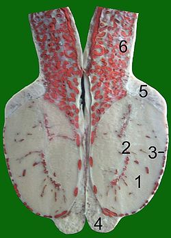
Section of a testicle of a bull, blood vessels injected with red gelatine. 1 parenchyma, 2 mediastinum testis, 3 tunica albuginea, 4 tail of epididymis, 5 head of epididymis, 6 spermatic cord with convoluted testicular artery
Details
Identifiers
Latin
tunica albuginea testis
FMA
19843
Anatomical terminology
[edit on Wikidata]
The tunica albuginea is a dense,[1][2] blue-white[3] layer of fibrous tissue surrounding the testis.[1][4] It is the middle of three envelopes forming the capsule of the testis; it is deep to the visceral layer of tunica vaginalis, and superficial to the tunica vasculosa testis (vascular layer of testis).[5]
The connective tissue of the tunica albuginea testis extends into the substance of the testis to form fibrous partitions - the septa testis.[1] At the posterior aspect of the testis (where the serosa of testis is deficient to allow for the attachment of the epididymis), the tunica albuginea extends into the testis to form the mediastinum testis.[5]
^ abcMartini, Frederic; Tallitsch, Robert B.; Nath, Judi L. (2017). Human Anatomy (9th ed.). Pearson. p. 711. ISBN 9780134320762.
^Standring, Susan (2020). Gray's Anatomy: The Anatomical Basis of Clinical Practice (42nd ed.). New York. p. 1292. ISBN 978-0-7020-7707-4. OCLC 1201341621.{{cite book}}: CS1 maint: location missing publisher (link)
^Standring, Susan (2020). Gray's Anatomy: The Anatomical Basis of Clinical Practice (42nd ed.). New York. p. 1292. ISBN 978-0-7020-7707-4. OCLC 1201341621.{{cite book}}: CS1 maint: location missing publisher (link)
^Federle, Michael P.; Rosado-de-Christenson, Melissa L.; Raman, Siva P.; Carter, Brett W., eds. (2017-01-01), "Testes and Scrotum", Imaging Anatomy: Chest, Abdomen, Pelvis (Second Edition), Elsevier, pp. 1000–1017, doi:10.1016/B978-0-323-47781-9.50043-X, ISBN 978-0-323-47781-9, retrieved 2021-02-03
^ abStandring, Susan (2020). Gray's Anatomy: The Anatomical Basis of Clinical Practice (42nd ed.). New York. p. 1292. ISBN 978-0-7020-7707-4. OCLC 1201341621.{{cite book}}: CS1 maint: location missing publisher (link)
and 27 Related for: Tunica albuginea of testis information
The tunicaalbuginea is a dense, blue-white layer of fibrous tissue surrounding the testis. It is the middle of three envelopes forming the capsule of the...
of the testis, with the tunicaalbugineaoftestis situated deep to it. Posteriorly, the visceral layer does not line the surface of the testis - instead...
mediastinum testis is a thick yet incomplete septum at the posterior part of the testis formed by the tunicaalbugineaoftestis projecting into the testis at...
of the digestive tract, lymph nodes, and some types of fascia. Other examples include periosteum and perichondrium of bones, and the tunicaalbuginea...
extensions of the tunicaalbuginea - the dense fibrous connective tissue surface covering of the testis - into the substance of the testis. The septa converge...
fibrous septa which extend between the mediastinum testis and the tunicaalbuginea, and consists of from one to three, or more, minute convoluted tubes...
testes are covered by a tough fibrous shell called the tunicaalbuginea. Under the tunicaalbuginea, the testes contain very fine-coiled tubes called seminiferous...
tear in the tunicaalbuginea resulting in extrusion of the testicular contents, including the seminiferous tubules. It is a rare complication of testicular...
superior aspect of the testis. Upon reaching the testis, the testicular artery divides into branches, which penetrate the tunicaalbuginea and arborize over...
converted into the tunicaalbuginea, thus excluding the surface epithelium from any part in the formation of the tissue of the testis. The cords of the central...
midline of the body, although even this forms from the fusion of paired structures in the embryo. Under a tough membranous shell, the tunicaalbuginea, the...
layer of connective tissue called the tunicaalbuginea. The corpora cavernosa are innervated by lesser and greater cavernous nerves and form most of the...
complications arising from suture fixation (and required breach of the tunicaalbuginea) like infarction and abscess formation, however this is not supported...
highly vascular. On the surface of the organ this tissue is much condensed, and forms a layer (tunicaalbuginea) composed of short connective-tissue fibers...
the ductus deferens; others pierce the back part of the tunicaalbuginea, and supply the substance of the testicle. The internal spermatic artery supplies...
of the dermis blends into the dense connective tissue forming the tunicaalbugineaof the corpus spongiosum behind the glans. The external lining with...
ligament and the round ligament of the uterus. The periphery of the testicles are converted into the tunicaalbuginea. Cords of the central mass run together...
navicularis) and ends in the tunicaalbugineaof the human penis. Externally it is attached to the frenulum which extends lower on the neck of the penis. The septum...
is a visible line or ridge of tissue that runs on the ventral (urethral) side of the human penis beginning from the base of the shaft and ending in the...
of scrotum or scrotal septum is an incomplete vertical wall (septum) that divides the scrotum into two compartments –each containing a single testis....
the class of transcription factors called homeobox genes are found in clusters named A, B, C, and D on four separate chromosomes. Expression of these proteins...
The perineal raphe is a visible line or ridge of tissue on the body that extends from the anus through the perineum to the scrotum (male) or the vulva...
The bulb of penis is the proximal/posterior end of the (unpaired median) corpus spongiosum. Together with the two crura (one crus on each side of the bulb)...
a carrier, the infectious proteins would still have to cross the blood-testis barrier to make transmission possible. Sperm were first observed in 1677...
of the clitoris is essentially similar to that of the penis except for the absence of a subalbugineal layer interposed between the tunicaalbuginea and...
 Global Information