This article may be too technical for most readers to understand. Please help improve it to make it understandable to non-experts, without removing the technical details.(February 2020) (Learn how and when to remove this message)
This article about biology may be excessively human-centric. Please improve coverage for other species and discuss this issue on the talk page. (Learn how and when to remove this message)
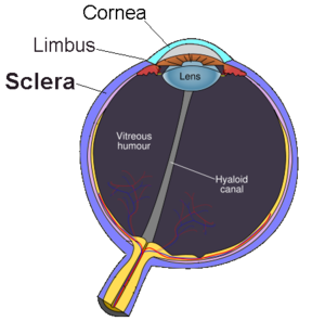
Sclera
The sclera, as separated from the cornea by the corneal limbus.
Details
Part of
Eye
System
Visual system
Artery
Anterior ciliary arteries, long posterior ciliary arteries, short posterior ciliary arteries
Identifiers
Latin
sclera
MeSH
D012590
TA98
A15.2.02.002
TA2
6750
FMA
58269
Anatomical terminology
[edit on Wikidata]
The sclera,[note 1] also known as the white of the eye or, in older literature, as the tunica albuginea oculi, is the opaque, fibrous, protective outer layer of the eye containing mainly collagen and some crucial elastic fiber.[2]
In the development of the embryo, the sclera is derived from the neural crest.[3] In children, it is thinner and shows some of the underlying pigment, appearing slightly blue. In the elderly, fatty deposits on the sclera can make it appear slightly yellow. People with dark skin can have naturally darkened sclerae, the result of melanin pigmentation.[4]
In humans, and some other vertebrates, the whole sclera is white or pale, contrasting with the coloured iris. The cooperative eye hypothesis suggests that the pale sclera evolved as a method of nonverbal communication that makes it easier for one individual to identify where another individual is looking. Other mammals with white or pale sclera include chimpanzees, many orangutans, some gorillas, and bonobos.[5]
^Mosby's Medical, Nursing, and Allied Health Dictionary (4th ed.). St. Louis: Mosby. 1994. p. 1402. ISBN 978-0815161134.
^Cassin, Barbara; Solomon, Sheila A.B. (1990). Dictionary of Eye Terminology (2nd ed.). Gainesville, Fla.: Triad Pub. Co. ISBN 978-0937404331.
^Hermann D. Schubert. Anatomy of the Orbit "New York Eye and Ear Infirmary of Mount Sinai - New York City - NYEE" (PDF). Archived from the original (PDF) on 2008-10-08. Retrieved 2008-05-19.
^Mukamal, Reena (30 July 2020). "Why Are the Whites of My Eyes Discolored?". American Academy of Ophthalmology. Retrieved 11 December 2020.
^Clark, Isabelle R.; Lee, Kevin C.; Poux, Tucker; Langergraber, Kevin E.; Mitani, John C.; Watts, David; Reed, James; Sandel, Aaron A. (2023-03-01). "White sclera is present in chimpanzees and other mammals". Journal of Human Evolution. 176: 103322. doi:10.1016/j.jhevol.2022.103322. ISSN 0047-2484. PMC 9998187. PMID 36706647. S2CID 256314941.
Cite error: There are <ref group=note> tags on this page, but the references will not show without a {{reflist|group=note}} template (see the help page).
The sclera, also known as the white of the eye or, in older literature, as the tunica albuginea oculi, is the opaque, fibrous, protective outer layer of...
Scleral tattooing is the practice of tattooing the sclera, or white part, of the human eye. Rather than being injected into the tissue, the dye is injected...
of the eye. It occurs due to weakening of outer layer of eye (cornea or sclera) by an inflammatory or degenerative condition. It may be of five types,...
spot of the sclera) is one of the ocular signs of death in which a reddish-brown discoloration is transversely arranged across the sclera. It occurs when...
English to refer to a folk belief according to which the visibility of the sclera above or under the irises has various meanings as an omen or symptom in...
The sclera and cornea form the fibrous tunic of the bulb of the eye; the sclera is opaque, and constitutes the posterior five-sixths of the tunic; the...
Joseph; Foster, C. Stephen (2012). "Noninflammatory Diseases of the Sclera". The Sclera. pp. 277–297. doi:10.1007/978-1-4419-6502-8_8. ISBN 978-1-4419-6501-1...
known as icterus, is a yellowish or greenish pigmentation of the skin and sclera due to high bilirubin levels. Jaundice in adults is typically a sign indicating...
eye. It contains connective tissues, and lies between the retina and the sclera. The human choroid is thickest at the far extreme rear of the eye (at 0...
with its outer layers, such as the outermost, white part of the eye (the sclera) and one of its inner layers (the pigmented choroid) keeping the eye essentially...
human cornea borders with the sclera at the corneal limbus. In lampreys, the cornea is solely an extension of the sclera, and is separate from the skin...
thin mucous membrane that lines the inside of the eyelids and covers the sclera (the white of the eye). It is composed of non-keratinized, stratified squamous...
known as a scleral contact lens, is a large contact lens that rests on the sclera and creates a tear-filled vault over the cornea. Scleral lenses are designed...
Trachypollia sclera is a species of sea snail, a marine gastropod mollusk in the family Muricidae, the murex snails or rock snails. Trachypollia sclera Woodring...
precisely between the inner retina and the outer fibrous layer composed of the sclera and cornea. The originally medieval Latin term comes from the Latin word...
forming the optic nerve exit the eye posteriorly through a hole in the sclera that is occupied by a mesh-like structure called the lamina cribrosa. It...
fatty layers of the bulbous conjunctiva and putting medications adjacent to sclera that is permeable to water, this will increase the penetration of the water-soluble...
limbus (Latin: corneal border) is the border between the cornea and the sclera (the white of the eye). It contains limbal stem cells in its palisades of...
The episclera is the outermost layer of the sclera (the white of the eye). It is composed of loose, fibrous, elastic tissue and attaches to Tenon's capsule...
obvious sign of hemolytic jaundice is the discolouration or yellowing of the sclera and the skin of the patient, but additional symptoms may be observed depending...
ciliary nerves then run forward and pierce the sclera at the back of the eye, traveling between the sclera and the choroid to innervate the iris sphincter...
the circle contact lens, which extend the appearance of the iris onto the sclera by having a dark tinted area all around. The result is an appearance of...
surgery, where an incision is made around the limbus, usually to expose the sclera and/or extraocular muscles for a variety of surgical procedures. "Peritomy"...
retinal detachment to indent or "buckle" the sclera inward, usually by sewing a piece of preserved sclera or silicone rubber to its surface. Laser photocoagulation...
white area of the eye, known as the sclera. The goal of this intervention is usually done to correct defects in sclera that resulted as a complication of...
diagnostic than examination of the bulbal conjunctiva, that overlying the sclera.[citation needed] Approximately 80% of cases of conjunctivitis in adults...
the eye. It is about 4 mm long, located near the junction of the iris and sclera, and is scalloped in appearance. The pars plana may not have a function...
 Global Information