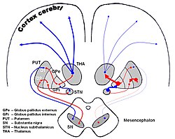
The image shows dopaminergic pathways of the human brain in normal condition (left) and Parkinsons Disease (right). Red Arrows indicate suppression of the target, blue arrows indicate stimulation of target structure. (Pallidothalamic connections visible but not labeled, as red line from GPi to THA.)
Anatomical terms of neuroanatomy
[edit on Wikidata]
The pallidothalamic tracts (or pallidothalamic connections)[1] are a part of the basal ganglia. They provide connectivity between the internal globus pallidus (GPi) and the thalamus, primarily the ventral anterior nucleus and the ventral lateral nucleus.
^Gallay MN, Jeanmonod D, Liu J, Morel A (August 2008). "Human pallidothalamic and cerebellothalamic tracts: anatomical basis for functional stereotactic neurosurgery". Brain Struct Funct. 212 (6): 443–63. doi:10.1007/s00429-007-0170-0. PMC 2494572. PMID 18193279.
and 7 Related for: Pallidothalamic tracts information
The pallidothalamictracts (or pallidothalamic connections) are a part of the basal ganglia. They provide connectivity between the internal globus pallidus...
field H3) is a large zone of mixed grey and white matter from the pallidothalamictracts of the lenticular fasciculus and the ansa lenticularis which combine...
cognitive and affective functions. It is mostly separated from the pallidothalamictracts. It can play a role in mediating symptoms in hereditary dystonia...
coming from different portions of the GPi. These tracts are collectively the pallidothalamictracts and join before they enter the ventral anterior nucleus...
retinohypothalamic tract (RHT) is a photic neural input pathway involved in the circadian rhythms of mammals. The origin of the retinohypothalamic tract is the intrinsically...
with field H1 of Forel. Nerve fibres form a tract containing cerebellothalamic (crossed) and pallidothalamic (uncrossed) fibres, that is situated between...
 Global Information