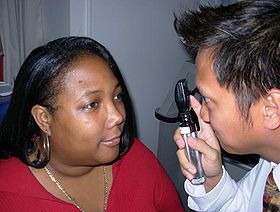
Ophthalmoscopic exam: the medical provider would next move in and observe with the ophthalmoscope from a distance of one to several cm.
MeSH
D009887
[edit on Wikidata]
Ophthalmoscopy, also called funduscopy, is a test that allows a health professional to see inside the fundus of the eye and other structures using an ophthalmoscope (or funduscope). It is done as part of an eye examination and may be done as part of a routine physical examination. It is crucial in determining the health of the retina, optic disc, and vitreous humor.[citation needed]
The pupil is a hole through which the eye's interior will be viewed. Opening the pupil wider (dilating it) is a simple and effective way to better see the structures behind it. Therefore, dilation of the pupil (mydriasis) is often accomplished with medicated eye drops before funduscopy. However, although dilated fundus examination is ideal, undilated examination is more convenient and is also helpful (albeit not as comprehensive), and it is the most common type in primary care.
An alternative or complement to ophthalmoscopy is to perform a fundus photography, where the image can be analysed later by a professional.
Ophthalmoscopy, also called funduscopy, is a test that allows a health professional to see inside the fundus of the eye and other structures using an ophthalmoscope...
Scanning laser ophthalmoscopy (SLO) is a method of examination of the eye. It uses the technique of confocal laser scanning microscopy for diagnostic imaging...
design of fundus cameras is based on the principle of monocular indirect ophthalmoscopy. A fundus camera provides an upright, magnified view of the fundus....
degeneration. The clinical macula is seen when viewed from the pupil, as in ophthalmoscopy or retinal photography. The term macula lutea comes from Latin macula...
directly observed in human subjects by adaptive optics scanning laser ophthalmoscopy, a real time imaging technique for examining retinal blood flow. The...
extraocular muscle motility, visual fields, intraocular pressure and ophthalmoscopy through a dilated pupil. A minimal eye examination consists of tests...
associated with the presence of changes in the optic fundus seen by ophthalmoscopy. The severity of the changes typical of hypertensive retinopathy is...
pressure, and is recommended in newly onset headaches. This may be done by ophthalmoscopy or fundus photography, and possibly slit lamp examination. It is important...
Optic disc Ophthalmoscopy photograph showing the optic disc as a bright area on the right where blood vessels converge. The terminal portion of the optic...
pepper syndrome. In 2006, Fahhad et al. published the results of an ophthalmoscopy study done on 4 children from 2 sibships of an Amish family; they showed...
variable corneal compensation (GDx VCC) and confocal scanning laser ophthalmoscopy (HRT II - Heidelberg Retina Tomograph). Also includes actual fundus...
being torn.[citation needed] A Weiss ring can sometimes be seen with ophthalmoscopy as very strong indicator that vitreous detachment has occurred. This...
wide range of applications, including optical imaging, microscopy, ophthalmoscopy, spectroscopy, and therapy. Examples of biomedical optics techniques...
pattern Hypertension with bradycardia and deteriorating consciousness Ophthalmoscopy for papilledema Bleeding diathesis (relative) Coagulopathy Decreased...
of the fundus of the eye. Once the pupil is dilated, examiners use ophthalmoscopy to view the eye's interior, which makes it easier to assess the retina...
Repka, Michael X.; Eghrari, Allen O. (2015), "Google glass indirect Ophthalmoscopy", MTM, 4 (1): 15–19, doi:10.7309/jmtm.4.1.4, archived from the original...
Jaxtthal; (Treatise on maladies of the fundus of the eye and an atlas of ophthalmoscopy). De l'iridotomie, 1873 (Iridotomy). Échelle métrique pour mesurer l'acuité...
and optic nerve. The portion of the posterior segment visible during ophthalmoscopy (or fundoscopy) is sometimes referred to as the posterior pole, or fundus...
difficult to take retinal images of animals with a tapetum lucidum because ophthalmoscopy devices designed for humans rely on a high level of on-axis illumination...
viticultural area in San Luis Obispo County, California, US Scanning laser ophthalmoscopy, an eye examination method Secondary lymphoid organ, for example a lymph...
B-scan Corneal topography Optical coherence tomography Scanning laser ophthalmoscopy Some of these techniques[example needed] are still at a research stage...
visualized by a slit lamp with a high positive lens or by using direct ophthalmoscopy; however, frequently there is no abnormal appearance of the nerve head...
extended associated with myopia (nearsightedness). It is diagnosed by ophthalmoscopy, which shows an area of retinal excavation in the region of the staphyloma...
available for the diagnosis of diseases and disorders affecting the retina. Ophthalmoscopy and fundus photography have long been used to examine the retina. Recently...
retinal correspondence A/V Arteriole–venue ratio BIO Binocular indirect ophthalmoscopy BSV Binocular single vision BV Binocular vision BVD Back vertex distance...
 Global Information