This article is about photography using microscopes. For images formed with tiny letters, see Micrography. For miniaturized photographs, see Microphotograph.
This article needs additional citations for verification. Please help improve this article by adding citations to reliable sources. Unsourced material may be challenged and removed. Find sources: "Micrograph" – news · newspapers · books · scholar · JSTOR(September 2014) (Learn how and when to remove this message)
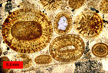
100x light micrograph of Meissner's corpuscle at the tip of a dermal papillus.40x micrograph of a canine rectum cross section.A photomicrograph of a thin section of a limestone with ooids. The largest is approximately 1.2 mm in diameter. The red object in the lower left is a scale bar indicating relative size.Approximately 10x micrograph of a doubled die on a coin, where the date was punched twice in the die used to strike the coin.
A micrograph or photomicrograph is a photograph or digital image taken through a microscope or similar device to show a magnified image of an object. This is opposed to a macrograph or photomacrograph, an image which is also taken on a microscope but is only slightly magnified, usually less than 10 times. Micrography is the practice or art of using microscopes to make photographs.
A micrograph contains extensive details of microstructure. A wealth of information can be obtained from a simple micrograph like behavior of the material under different conditions, the phases found in the system, failure analysis, grain size estimation, elemental analysis and so on. Micrographs are widely used in all fields of microscopy.
A micrograph or photomicrograph is a photograph or digital image taken through a microscope or similar device to show a magnified image of an object. This...
and map their distribution. Due to the very narrow electron beam, SEM micrographs have a large depth of field yielding a characteristic three-dimensional...
immature male analog, the immature glans penis. Micrograph of the primordial phallus, H&E stain. Micrograph of the primordial phallus, H&E stain. Aphallia...
5 cm long Intermediate magnification micrograph of a Leydig cell tumour, H&E stain High magnification micrograph of a Leydig cell tumour, H&E stain Cross-section...
magnification micrograph of a molluscum contagiosum lesion Low-magnification micrograph of molluscum contagiosum, H&E stain High-magnification micrograph of molluscum...
of the gallbladder). Micrograph of cholesterolosis of the gallbladder Micrograph of cholesterolosis of the gallbladder Micrograph of cholesterolosis of...
R‑banding. Staining with Giemsa confers a purple color to chromosomes, but micrographs are often converted to grayscale to facilitate data presentation and...
2012 A. wuliandei Zhu et al. 2020 Micrograph of actinomycosis, H&E stain Micrograph of actinomycosis, GMS stain Micrograph of actinomycosis, Gram stain Harz...
adenocarcinoma from biopsy. H&E stain. Micrograph of decidualized endometrium due to exogenous progesterone. H&E stain. Micrograph of decidualized endometrium due...
melanin (rare). Micrograph showing melanosis coli, which appears as brown pigmentation in the macrophages in the lamina propria. Micrograph of melanosis...
provided a comprehensive description of them in 1903. High magnification micrograph of a Brenner tumor showing the characteristic coffee bean nuclei which...
needed] Low magnification micrograph of an SSL. Intermediate magnification micrograph of an SSL. High magnification micrograph of a SSL showing crypt branching...
Sapporo virus Transmission electron micrograph of Sapporo viruses Virus classification (unranked): Virus Realm: Riboviria Kingdom: Orthornavirae Phylum:...
Granulomatous diseases Sarcoidosis Micrograph of asteroid bodies in pulmonary sarcoidosis. H&E stain. Micrograph of asteroid bodies in pulmonary sarcoidosis...
histopathologic finding of steatohepatitis. Micrograph showing a Mallory body. Original magnification 400X. H&E stain. Micrograph showing a Mallory body. Original...
Medium-power magnification micrograph of a H&E stained slide showing a portion of a vaginal wall. Stratified squamous epithelium and underling connective...
connected lobes that stay in the cell until the end of its cell life. Micrograph of an infarct in the biliary tract, with pyknotic nuclei (arrows) (400x)...
A fluorescence microscope is an optical microscope that uses fluorescence instead of, or in addition to, scattering, reflection, and attenuation or absorption...
 Global Information