This article includes a list of general references, but it lacks sufficient corresponding inline citations. Please help to improve this article by introducing more precise citations.(May 2015) (Learn how and when to remove this message)
Hyaloid artery
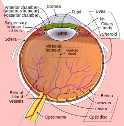
The hyaloid artery is located on the gray band near the center of the image.
Details
Source
central retinal artery
Identifiers
Latin
arteria hyaloidea
TA98
A15.2.06.009
TA2
6812
FMA
77670
Anatomical terminology
[edit on Wikidata]
The hyaloid artery is a branch of the ophthalmic artery, which is itself a branch of the internal carotid artery. It is contained within the optic stalk of the eye and extends from the optic disc through the vitreous humor to the lens. Usually fully regressed before birth, its purpose is to supply nutrients to the developing lens in the growing fetus.
During the tenth week of development in humans (time varies depending on species), the lens grows independent of a blood supply and the hyaloid artery usually regresses. Its proximal portion remains as the central artery of the retina. Regression of the hyaloid artery leaves a clear central zone through the vitreous humor, called the hyaloid canal or Cloquet's canal. Cloquet's canal is named after the French physician Jules Germain Cloquet (1790–1883) who first described it.
Occasionally the artery may not fully regress, resulting in the condition persistent hyaloid artery. More commonly, small remnants of the artery may remain. Free remnants can sometimes be seen as "floaters". An anterior remnant of the hyaloid artery can be seen in some people as Mittendorf's dot, a small pinpoint-like scar on the posterior surface of the lens.[1] A posterior remnant may be seen where the artery left the optic disc, and is known as Bergmeister's papilla.
^Lee Ann Remington, Clinical Anatomy of the Visual System, 2005 p. 124
The hyaloidartery is a branch of the ophthalmic artery, which is itself a branch of the internal carotid artery. It is contained within the optic stalk...
invagination of the hyaloid membrane, which encloses the vitreous body. In the fetus, the hyaloid canal contains a prolongation of the central artery of the retina...
have had inflammation inside the eye.[better source needed] The hyaloidartery, an artery running through the vitreous humour during the fetal stage of...
posterior lens capsule, which represents the site of attachment of the hyaloidartery before it subsequently regressed. Bergmeister's papilla: A tuft of fibrous...
case of central retinal artery occlusion (CRAO). The central retinal artery is formed from the proximal part of the hyaloidartery after atrophy of its distal...
or retinal detachment, liquefaction of the vitreous and a persistent hyaloidartery. The malformations of the retina are dominant (i.e. they occur in heterozygous...
eyes with mostly hyaloidartery changes Mittendorf dot: the termination of the normally regressed anterior portion of the hyaloidartery, found slightly...
railroad Cloquet Valley State Forest, Minnesota Cloquet's canal, see Hyaloidartery Cloquet's node, a lymph node This disambiguation page lists articles...
also the first to describe and identify the remnant of the embryonic hyaloidartery. This vestige was to become known as Cloquet's canal. Cloquet's name...
that surrounds the lens. It receives arterial blood supply from the hyaloidartery. This blood supply slowly regress and vascular capsule disappear before...
response to the raised pressure: subhyaloid hemorrhage (bleeding under the hyaloid membrane, which envelops the vitreous body of the eye) and vitreous hemorrhage...
eye resulting from failure of the embryological, primary vitreous, and hyaloid vasculature to regress, whereby the eye is shorter, develops a cataract...
segment is the back five-sixths of the eye that includes the anterior hyaloid membrane and all of the optical structures behind it: the vitreous humor...
physical activities. Valsalva retinopathy is a form of sub-retinal, sub-hyaloid or sub-internal limiting membrane hemorrhage occur due to rupture of retinal...
 Global Information