Small pit in the retina of the eye responsible for all central vision
"Central fovea" redirects here. For other uses, see Fovea (disambiguation).
Not to be confused with the optic disc, a nearby structure that also carries signals to the optic nerve.
Fovea centralis
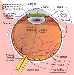
Schematic diagram of the human eye, with the fovea at the bottom. It shows a horizontal section through the right eye.
Details
Identifiers
Latin
fovea centralis
MeSH
D005584
TA98
A15.2.04.022
TA2
6785
FMA
58658
Anatomical terminology
[edit on Wikidata]
The fovea centralis is a small, central pit composed of closely packed cones in the eye. It is located in the center of the macula lutea of the retina.[1][2]
The fovea is responsible for sharp central vision (also called foveal vision), which is necessary in humans for activities for which visual detail is of primary importance, such as reading and driving. The fovea is surrounded by the parafovea belt and the perifovea outer region.[2]
The parafovea is the intermediate belt, where the ganglion cell layer is composed of more than five layers of cells, as well as the highest density of cones; the perifovea is the outermost region where the ganglion cell layer contains two to four layers of cells, and is where visual acuity is below the optimum. The perifovea contains an even more diminished density of cones, having 12 per 100 micrometres versus 50 per 100 micrometres in the most central fovea. That, in turn, is surrounded by a larger peripheral area, which delivers highly compressed information of low resolution following the pattern of compression in foveated imaging.[citation needed]
Approximately half the nerve fibers in the optic nerve carry information from the fovea, while the remaining half carry information from the rest of the retina. The parafovea extends to a radius of 1.25 mm from the central fovea, and the ':perifovea is found at a 2.75 mm radius from the fovea centralis.[3]
The term fovea comes from Latin fovea 'pit'.
The fovea centralis was named by German histologist Carl Bergmann.[4]
^"Simple Anatomy of the Retina". Webvision. University of Utah. Archived from the original on 2011-03-15. Retrieved 2011-09-28.
^ abIwasaki, M; Inomata, H (1986). "Relation between superficial capillaries and foveal structures in the human retina". Investigative Ophthalmology & Visual Science. 27 (12): 1698–705. PMID 3793399.
^"eye, human."Encyclopædia Britannica. 2008. Encyclopædia Britannica 2006 Ultimate Reference Suite DVD
^Thibos, Larry; Lenner, Katharina; Thibos, Cameron (18 Dec 2023). "Carl Bergmann (1814–1865) and the discovery of the anatomical site in the retina where vision is initiated". Journal of the History of the Neurosciences. 33 (2): 180–203. doi:10.1080/0964704X.2023.2286991. PMID 38109332. S2CID 266361309.
The foveacentralis is a small, central pit composed of closely packed cones in the eye. It is located in the center of the macula lutea of the retina...
structure. Foveacentralis of the retina Fovea buccalis or dimple Fovea of the femoral head Trochlear fovea of the frontal bone Pterygoid fovea of the mandible...
The foveal avascular zone (FAZ) is a region within the foveacentralis at the centre of the retina of the human eye that is devoid of retinal blood vessels...
light, or the scotopic region. Cone cells are densely packed in the foveacentralis, a 0.3 mm diameter rod-free area with very thin, densely packed cones...
"center" of the retina (the point directly behind the lens) lies the fovea (or foveacentralis), which contains only cone cells; and is the region capable of...
sensitivity of the cones it is impossible to discriminate colors. In the foveacentralis, cones predominate and are present at high density. The macula is thus...
central retina adapted for high-acuity vision. This area, termed the foveacentralis, is avascular (does not have blood vessels), and has minimal neural...
have a small area of the retina with very high visual acuity, the foveacentralis. It covers about 2 degrees of visual angle in people. To get a clear...
confined to a single or a few sources of leakage at a safe distance from the fovea. Laser photocoagulation is not indicated for cases where the leak is very...
attempting to produce an image from a human eye is that the size of the foveacentralis, the actual focal point of the image on the retina, is very small (about...
(1999). "Müller Cell Cone, an Overlooked Part of the Anatomy of the FoveaCentralis". Archives of Ophthalmology. 117 (6): 821–3. doi:10.1001/archopht.117...
of different breeds can have different retina configurations. The foveacentralis area of dogs' eyes, which is attached to a nerve fiber, is the most...
epithelium hypertrophy and impacted teeth. Bruch's membrane Drusen Foveacentralis Fundus (eye) Macula of retina This article incorporates text in the...
primates, move their entire eyes to focus images of interest onto their foveacentralis. In jumping spiders with a translucent carapace, such movements within...
A Palaeozoic Geology of London, Ontario (1974), Coach House Press FoveaCentralis (1975), Coach House Press Alter Sublime (1980), Coach House Press Predators...
PMID 22947032. "Acanthogonatus centralis". Integrated Taxonomic Information System. ADW entry "Acanthogonatus centralis" at the Encyclopedia of Life ZipcodeZoo...
circular area called the macula. The center of this circular area is the fovea. The fovea and a small area surrounding it are not supplied by the central retinal...
and it has a procurved fovea. Its labium possesses no cuspules. A serrula is present. Its sternal sigilla is as in A. Centralis, and it has a lightly reborder...
some species. Towards the centre of the retina is the fovea (or the less specialised, area centralis) which has a greater density of receptors and is the...
of pathologies affecting the central part of the retina (macula and foveacentralis) as patients with these pathologies are often unable to fixate reliably...
females are recognized by the spermathecae (similar to A. fuegianus, A. centralis and A. parana, which have - unlike A. Confusus - no inferior tarsal claws...
Although they lack a fovea, some diurnal lemurs have a cone-rich, although less clustered, area centralis. This area centralis has a high rod-to-cone...
 Global Information