Global Information Lookup Global Information

Echocardiography information


Echocardiography
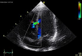
An abnormal echocardiogram: Image shows a midmuscular ventricular septal defect. The trace in the lower left shows the cardiac cycle and the red mark the time in the cardiac cycle when the image was captured. Colors are used to represent the velocity and direction of blood flow.
ICD-9-CM88.72
MeSHD004452
OPS-301 code3-052
MedlinePlus003869
[edit on Wikidata]

Echocardiography, also known as cardiac ultrasound, is the use of ultrasound to examine the heart. It is a type of medical imaging, using standard ultrasound or Doppler ultrasound.[1] The visual image formed using this technique is called an echocardiogram, a cardiac echo, or simply an echo.

Echocardiography is routinely used in the diagnosis, management, and follow-up of patients with any suspected or known heart diseases. It is one of the most widely used diagnostic imaging modalities in cardiology. It can provide a wealth of helpful information, including the size and shape of the heart (internal chamber size quantification), pumping capacity, location and extent of any tissue damage, and assessment of valves. An echocardiogram can also give physicians other estimates of heart function, such as a calculation of the cardiac output, ejection fraction, and diastolic function (how well the heart relaxes).

Echocardiography is an important tool in assessing wall motion abnormality in patients with suspected cardiac disease. It is a tool which helps in reaching an early diagnosis of myocardial infarction, showing regional wall motion abnormality. Also, it is important in treatment and follow-up in patients with heart failure, by assessing ejection fraction.[2][3]

Echocardiography can help detect cardiomyopathies, such as hypertrophic cardiomyopathy, and dilated cardiomyopathy. The use of stress echocardiography may also help determine whether any chest pain or associated symptoms are related to heart disease.

The most important advantages of echocardiography are that it is not invasive (does not involve breaking the skin or entering body cavities) and has no known risks or side effects.[4]

Not only can an echocardiogram create ultrasound images of heart structures, but it can also produce accurate assessment of the blood flowing through the heart by Doppler echocardiography, using pulsed- or continuous-wave Doppler ultrasound. This allows assessment of both normal and abnormal blood flow through the heart. Color Doppler, as well as spectral Doppler, is used to visualize any abnormal communications between the left and right sides of the heart, any leaking of blood through the valves (valvular regurgitation), and estimate how well the valves open (or do not open in the case of valvular stenosis). The Doppler technique can also be used for tissue motion and velocity measurement, by tissue Doppler echocardiography.

Echocardiography was also the first ultrasound subspecialty to use intravenous contrast. Echocardiography is performed by cardiac sonographers, cardiac physiologists (UK), or physicians trained in echocardiography.

Recognized as the "Father of Echocardiography", the Swedish physician Inge Edler (1911–2001), a graduate of Lund University, was the first of his profession to apply ultrasonic pulse echo imaging in diagnosing cardiac disease, which the acoustical physicist Floyd Firestone had developed to detect defects in metal castings. In fact, Edler in 1953 produced the first echocardiographs using an industrial Firestone-Sperry Ultrasonic Reflectoscope. In developing echocardiography, Edler worked with the physicist Carl Hellmuth Hertz, the son of the Nobel laureate Gustav Hertz and grandnephew of Heinrich Rudolph Hertz.[5][6]

  1. ^ Cleve, Jayne; McCulloch, Marti L. (2018), Nihoyannopoulos, Petros; Kisslo, Joseph (eds.), "Conducting a Cardiac Ultrasound Examination", Echocardiography, Springer International Publishing, pp. 33–42, doi:10.1007/978-3-319-71617-6_2, ISBN 978-3319716176
  2. ^ Oh, J. K. (1 January 2007). "Echocardiography in heart failure: Beyond diagnosis". European Journal of Echocardiography. 8 (1): 4–14. doi:10.1016/j.euje.2006.09.002. ISSN 1525-2167. PMID 17240313.
  3. ^ Modin, Daniel; Andersen, Ditte Madsen; Biering-Sørensen, Tor (June 2018). "Echo and heart failure: when do people need an echo, and when do they need natriuretic peptides?". Echo Research and Practice. 5 (2): R65–R79. doi:10.1530/erp-18-0004. PMC 5958420. PMID 29691224.
  4. ^ Hanton, G.; Eder, V.; Rochefort, G.; Bonnet, P.; Hyvelin, J. M. (2008). "Echocardiography, a non-invasive method for the assessment of cardiac function and morphology in preclinical drug toxicology and safety pharmacology". Expert Opinion on Drug Metabolism & Toxicology. 4 (6): 681–696. doi:10.1517/17425255.4.6.681. PMID 18611111. S2CID 72290828. Retrieved 30 June 2021.
  5. ^ Batohi, Bhavna; Sidhu, Paul S. (2014). "The Development of Ultrasound for Clinical Use". In Thompson, Gilbert (ed.). Pioneers of Medicine Without a Nobel Prize. World Scientific. pp. 141–159. ISBN 978-1783263868. Retrieved 23 September 2016.
  6. ^ Singh, Siddharth; Goyal, Abha (2007). "The origin of echocardiography: A Tribute to Inge Edler". Tex. Heart Inst. J. 34 (4): 431–438. PMC 2170493. PMID 18172524.

and 29 Related for: Echocardiography information

Request time (Page generated in 0.558 seconds.)

Echocardiography

Last Update:

Echocardiography, also known as cardiac ultrasound, is the use of ultrasound to examine the heart. It is a type of medical imaging, using standard ultrasound...

Word Count : 3997

Transesophageal echocardiogram

Last Update:

professional organizations recommend against using transesophageal echocardiography to detect cardiac sources of embolization after a patient's health...

Word Count : 2373

Doppler echocardiography

Last Update:

Doppler echocardiography is a procedure that uses Doppler ultrasonography to examine the heart. An echocardiogram uses high frequency sound waves to create...

Word Count : 2290

Cardiac stress test

Last Update:

narrowed (~70% or more). A stress test may be accompanied by echocardiography. The echocardiography is performed both before and after the exercise so that...

Word Count : 3372

Intracardiac echocardiogram

Last Update:

Intracardiac echocardiography (ICE) is a specialized form of echocardiography that utilizes an ultrasound-tipped catheter to perform imaging of the heart...

Word Count : 543

Fetal echocardiography

Last Update:

Fetal echocardiography, or Fetal echocardiogram, is the name of the test used to diagnose cardiac conditions in the fetal stage. Cardiac defects are amongst...

Word Count : 935

Speckle tracking echocardiography

Last Update:

In the fields of cardiology and medical imaging, speckle tracking echocardiography (STE) is an echocardiographic imaging technique. It analyzes the motion...

Word Count : 1991

Tissue Doppler echocardiography

Last Update:

Tissue Doppler echocardiography (TDE) is a medical ultrasound technology, specifically a form of echocardiography that measures the velocity of the heart...

Word Count : 2665

Strain rate imaging

Last Update:

Strain rate imaging is a method in echocardiography (medical ultrasound) for measuring regional or global deformation of the myocardium (heart muscle)...

Word Count : 3328

Left ventricular hypertrophy

Last Update:

include OGN, osteoglycin. The commonly used method to diagnose LVH is echocardiography, with which the thickness of the muscle of the heart can be measured...

Word Count : 1442

American Society of Echocardiography

Last Update:

of Echocardiography (ASE) is a professional organization of physicians, cardiac sonographers, nurses and scientists involved in echocardiography, the...

Word Count : 204

Intravascular ultrasound

Last Update:

Intravascular ultrasound (IVUS) or intravascular echocardiography is a medical imaging methodology using a specially designed catheter with a miniaturized...

Word Count : 2464

Heart failure

Last Update:

heart to pump. Diagnosis is based on symptoms, physical findings, and echocardiography. Blood tests, and a chest x-ray may be useful to determine the underlying...

Word Count : 15558

Numerical modeling in echocardiography

Last Update:

Numerical manipulation of Doppler parameters obtain during routine Echocardiography has been extensively utilized to non-invasively estimate intra-cardiac...

Word Count : 385

Cardiothoracic anesthesiology

Last Update:

transesophageal echocardiography, intraspinal drainage placement, and advanced ultrasound guidance of vascular access.[citation needed] Echocardiography produces...

Word Count : 1698

Focus assessed transthoracic echocardiography

Last Update:

Focus assessed transthoracic echocardiography (or FATE) is a type of transthoracic echocardiogram, or sonogram of the heart, often performed by non-cardiologist...

Word Count : 135

Pulmonary embolism

Last Update:

submassive PE, dysfunction of the right side of the heart may be seen on echocardiography, an indication that the pulmonary artery is severely obstructed and...

Word Count : 9695

Medical ultrasound

Last Update:

modalities, or by cardiologists in the case of cardiac ultrasonography (echocardiography). Sonography is effective for imaging soft tissues of the body. Superficial...

Word Count : 13632

Medical imaging

Last Update:

is used to image the heart it is referred to as an echocardiogram. Echocardiography allows detailed structures of the heart, including chamber size, heart...

Word Count : 7144

Pulmonary hypertension

Last Update:

based on the above assessments, echocardiography is performed as the next step. A meta-analysis of Doppler echocardiography for predicting the results of...

Word Count : 8891

Transthoracic echocardiogram

Last Update:

abbreviated "TTE", it can be easily confused with transesophageal echocardiography which is abbreviated "TEE". Pronunciation of "TTE" and "TEE" are similar...

Word Count : 3161

Ejection fraction

Last Update:

computational applications. The first common measurement method is echocardiography, although cardiac magnetic resonance imaging (MRI), cardiac computed...

Word Count : 2219

Atrial septal defect

Last Update:

normally closed, but can open under increased right atrial pressure. On echocardiography, shunting of blood may not be noted except when the patient coughs...

Word Count : 6230

Mitral valve

Last Update:

valve. This early filling across the mitral valve is seen on doppler echocardiography of the mitral valve as the E wave. After the E wave, there is a period...

Word Count : 2337

Vena contracta

Last Update:

primary pipe flow). Measurement of the vena contracta is useful in echocardiography, where it describes the smallest area of the blood flow jet as it exits...

Word Count : 547

Abdominal guarding

Last Update:

Abdominal guarding is the tensing of the abdominal wall muscles to guard inflamed organs within the abdomen from the pain of pressure upon them. The tensing...

Word Count : 449

Pulmonary edema

Last Update:

diseases. Especially in the case of cardiogenic pulmonary edema, urgent echocardiography may strengthen the diagnosis by demonstrating impaired left ventricular...

Word Count : 3664

Cardiac catheterization

Last Update:

coronary sinus (for the left ventricle stimulation).[citation needed] Echocardiography is a non-invasive method to evaluate the heart valves. However, sometimes...

Word Count : 2876

Sonographer

Last Update:

A sonographer performing pediatric echocardiography...

Word Count : 2262

PDF Search Engine © AllGlobal.net