Brainstem (basilar part of pons not labeled, but is visible at bottom)
Details
Identifiers
Latin
Pars basilaris pontis, basis pontis
NeuroNames
616
NeuroLex ID
birnlex_1043
TA98
A14.1.05.101
TA2
5925
FMA
72244
Anatomical terms of neuroanatomy
[edit on Wikidata]
The basilar part of pons, also known as basis pontis, is the ventral part of the pons; the dorsal part is known as the pontine tegmentum.
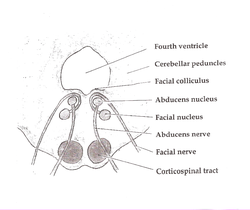
The basilar pons makes up two thirds of the pons within the brainstem.[1] It has a ridged appearance with a shallow groove at the midline. This groove is called the basilar sulcus and is covered by the basilar artery,[2] which feeds into the Circle of Willis and provides blood supply to the brainstem and cerebellum.[3] The basilar pons has this kind of appearance due to the fibers that come out of the pons and enter the cerebellum.[2] This part of the brainstem contains fibers from the corticospinal tract (a descending pathway for neurons to reach other structures in the body), pontine nuclei, and transverse pontine fibers.[1] The corticospinal tract carries neurons from the primary motor cortex in the brain to the spinal cord, aiding in voluntary motor movement of the body. In addition to passing through the basilar pons, corticospinal tract fibers go through other structures of the brainstem, such as the internal capsule and the crus cerebri.[4]
An integral part of the basilar pons is the pontine nuclei. The pontine nuclei are responsible for projecting axons that go to the opposite cerebellar hemisphere through the middle cerebellar peduncle. Doing this makes the axons change into the transverse pontine fibers.[1] The fibers of the pontine nuclei are all important to motor function, including fiber bundles such as the corticospinal fibers and corticopontine-pontocerebellar system.[5] Specifically, the basilar pons contains all the corticofugal fibers, which include the corticospinal, corticobulbar (or corticonuclear), and corticopontine fibers.[6] The basal pontine nuclei provides the most information to the cerebellum. These pontine nuclei are integral in helping the basilar pons carry information from the cerebral cortex to the cerebellum. The basilar pons is able to do this via the corticopontine fibers that it receives. Once the information passes from the cerebral cortex to the basilar pons and then finally to the cerebellum, the cerebellum gets information regarding complex cognitive functions.[7]
^ abcJohns, P. (2014). Clinical Neuroscience. Elsevier Health Sciences. pp. 27–47.
^ abMichael-Titus, A. (2010). The Nervous System: Second Edition. Churchill Livingstone.
^Adigun, O. (2020). Anatomy, Head and Neck, Basilar Artery. StatPearls Publishing. PMID 29083786.
^Gould, D.J. (2016). Nolte's the Human Brain: An Introduction to its Functional Anatomy. Elsevier Health Sciences.
^Mihailoff, G.A.; Haines, D.E. (2018), "The Pons and Cerebellum", Fundamental Neuroscience for Basic and Clinical Applications, Elsevier, pp. 172–182, doi:10.1016/b978-0-323-39632-5.00012-8, ISBN 978-0-323-39632-5
^Schmahmann, Jeremy D.; Pandya, Deepak N. (October 1995). "Prefrontal cortex projections to the basilar pons in rhesus monkey: implications for the cerebellar contribution to higher function". Neuroscience Letters. 199 (3): 175–178. doi:10.1016/0304-3940(95)12056-A. PMID 8577391. S2CID 42572383.
and 28 Related for: Basilar part of pons information
The basilarpartofpons, also known as basis pontis, is the ventral partof the pons; the dorsal part is known as the pontine tegmentum. The basilar pons...
The basilar sulcus (groove for basilar artery) is a groove in the pons, partof the brainstem. The basilar sulcus is vertical directed and lies in the...
The pons (pl.: pontes; from Latin pons, "bridge") is partof the brainstem that in humans and other mammals, lies inferior to the midbrain, superior to...
(CN VI). It ascends along the basilar sulcus of the ventral pons. It divides at the junction of the midbrain and pons into the posterior cerebral arteries...
part of occipital bone Basilar partofponsBasilar plexus Basilar sinus Basilar skull fracture Basilar sulcus of the pons This disambiguation page lists...
constituents of the circle of Willis, cerebellar arteries, and basilar artery". Other lesions that are associated with lacunes appear in the "deep nuclei of the...
dorsal pons, is located within the brainstem, and is one of two parts of the pons, the other being the ventral pons or basilarpartof the pons. The pontine...
form the basilar artery at the base of the pons. The basilar artery is the main blood supply to the brainstem and connects to the Circle of Willis to...
(AICA) is one of three pairs of arteries that supplies blood to the cerebellum. It arises from the basilar artery on each side at the level of the junction...
from the basilar artery. It conveys information from the cerebrum and the pons to the cerebellum. The middle cerebellar peduncle is the largest of the three...
center. The pons co-ordinates activities of the cerebellar hemispheres. The pons and medulla oblongata are parts of the hindbrain that form much of the brainstem...
inferior petrosal sulcus, formed by the junction of the petrous partof the temporal bone with the basilarpartof the occipital bone. It begins below and behind...
portion of the basilar artery, just inferior to its bifurcation into the posterior cerebral artery. Here, it wraps posteriorly around the pons (to which...
or posteromedial central arteries, are pontine arteries – branches of the basilar artery that supply the pontine nuclei, corticobulbar tract, corticospinal...
propagation properties of the cochlear partition The Organ of Corti, the sensory epithelium, a cellular layer on the basilar membrane, in which sensory...
basilar portion of the occipital bone; it supports the upper partof the pons. The lateral surfaces of the body are united with the greater wings of the...
runs between the base of the pons and medulla oblongata (the lower portion of the brainstem). This junction between the pons, medulla, and cerebellum that...
caused by the displacement of the brainstem stretching and tearing perforating branches of the basilar artery to the pons; venous infarction may play...
motor), and hair bundles to shift which, in turn, electrically affects the basilar membrane's movement (hair-bundle motor). These motors (outer hair cells)...
junction of the petrous partof the temporal bone of the skull with the basilarpartof the occipital bone. It begins in the postero-inferior partof the cavernous...
OHC damage and thus a loss of sensitivity to quiet sounds, occurs more than IHC loss. When the IHCs or partof the basilar membrane are damaged or destroyed...
condition involving severe damage to the myelin sheath of nerve cells in the pons (an area of the brainstem). It is predominately iatrogenic (treatment-induced)...
parahippocampal gyrus and anterior partof the lingual gyrus of the right hemisphere). The opposite of prosopagnosia is the skill of superior face recognition...
mesencephalon is the rostral-most portion of the brainstem connecting the diencephalon and cerebrum with the pons. It consists of the cerebral peduncles, tegmentum...
the caudal portion of the pons near the midline, medial to the sulcus limitans. The abducens nucleus along with the internal genu of the facial nerve make...
 Global Information