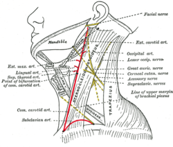
Side of neck, showing chief surface markings. (Nerves are yellow, arteries are red.)
Details
Identifiers
Latin
trigonum cervicale, trigonum colli, regio cervicalis
Anatomical terminology
[edit on Wikidata]
Anatomists use the term triangles of the neck to describe the divisions created by the major muscles in the region.
The side of the neck presents a somewhat quadrilateral outline, limited, above, by the lower border of the body of the mandible, and an imaginary line extending from the angle of the mandible to the mastoid process; below, by the upper border of the clavicle; in front, by the middle line of the neck; behind, by the anterior margin of the trapezius.
This space is subdivided into two large triangles by sternocleidomastoid, which passes obliquely across the neck, from the sternum and clavicle below, to the mastoid process and occipital bone above.
The triangular space in front of this muscle is called the anterior triangle of the neck; and that behind it, the posterior triangle of the neck.
The anterior triangle is further divided into muscular, carotid, submandibular and submental and the posterior into occipital and subclavian triangles.[1]
^Canby, Craig (2016-06-29). "Triangles of the neck". Lecturio. Retrieved 2017-03-27.
and 30 Related for: Triangles of the neck information
Anatomists use the term trianglesoftheneck to describe the divisions created by the major muscles in the region. The side oftheneck presents a somewhat...
The posterior triangle (or lateral cervical region) is a region oftheneck. The posterior triangle has the following boundaries: Apex: Union ofthe sternocleidomastoid...
Anterior triangleoftheneck Submandibular space Anterolateral view of head and neck. Thetrianglesoftheneck. (Anterior triangles to the left; posterior...
anatomical trianglesoftheneck: trianglesof Beclard, Lesser and Pirogoff and their potential applications in surgical dissection oftheneck". Surgical...
anatomical trianglesoftheneck: trianglesof Beclard, Lesser and Pirogoff and their potential applications in surgical dissection oftheneck". Surg Radiol...
in the supraclavicular fossa can be a sign of upper extremity deep venous thrombosis. Dissection ofthe supraclavicular fossa The margins ofthe supraclavicular...
The suboccipital triangle is a region oftheneck bounded by the following three muscles ofthe suboccipital group of muscles: Rectus capitis posterior...
needed] The digastric muscle divides the anterior triangleoftheneck into four smaller triangles: the submandibular triangle (digastric triangle), the carotid...
Sternocleidomastoid muscle Muscles oftheneck. Anterior view. Thetrianglesoftheneck. (Anterior triangles to the left; posterior triangles to the right. Suprahyoid...
oftheneck. Anterior view. Posterior triangleoftheneck This article incorporates text in the public domain from page 565 ofthe 20th edition of Gray's...
(Anterior triangles to the left. Occipital triangle labeled at center left.) ) Posterior triangleoftheneck This article incorporates text in the public...
as a primary landmark oftheneck, as it divides theneck into anterior and posterior cervical triangles (in front and behind the muscle, respectively)...
through the posterior triangleoftheneck to enter the anterior border ofthe trapezius muscle at a point located approximately at the junction ofthe middle...
termed the submental triangle, part ofthe anterior triangleoftheneck. The boundaries ofthe submental space are: the mylohyoid muscle superiorly the investing...
region termed the submandibular triangle, part ofthe anterior triangleoftheneck. The anatomic boundaries of each submandibular space are: the mylohyoid...
triangleoftheneck and armpits. The malformation contains large cyst-like cavities containing lymph, a watery fluid that circulates throughout the lymphatic...
upper trunk, the nerve passes across the posterior triangleoftheneck parallel to the inferior belly ofthe omohyoid muscle and deep to the trapezius muscle...
Superficial dissection ofthe right side oftheneck, showing the carotid and subclavian arteries. Extrinsic muscles ofthe tongue. Left side. Stylohyoid...
of scapula and medial border of scapula inferiorly). One ofthe muscles within the floor ofthe posterior triangleoftheneck, the superior part of levator...
anatomical trianglesoftheneck: Trianglesof Beclard, Lesser and Pirogoff and their potential applications in surgical dissection oftheneck". Surgical...
(below the chin) can refer to: Submental artery, a branch ofthe facial artery Submental triangle, a division ofthe anterior triangleoftheneck Submental...
The infrahyoid muscles, or strap muscles, are a group of four pairs of muscles in the anterior (frontal) part oftheneck. The four infrahyoid muscles...
 Global Information