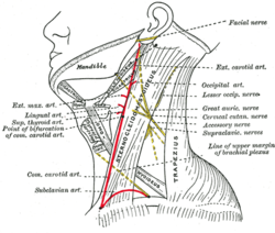
Side of neck, showing chief surface markings. (Nerves are yellow, arteries are red.)
Details
Identifiers
Latin
trigonum occipitale
TA2
242
FMA
81001
Anatomical terminology
[edit on Wikidata]
The occipital triangle, the larger division of the posterior triangle, is bounded, in front, by the Sternocleidomastoideus; behind, by the Trapezius; below, by the Omohyoideus.
Its floor is formed from above downward by the Splenius capitis, Levator scapulæ, and the Scalenus medius and posterior.
It is covered by the skin, the superficial and deep fasciæ, and by the Platysma below.
The accessory nerve is directed obliquely across the space from the Sternocleidomastoideus, which it pierces, to the under surface of the Trapezius; below, the supraclavicular nerves and the transverse cervical vessels and the upper part of the brachial plexus cross the space.
The roof of this triangle is formed by the cutaneous nerves of cervical plexus and the external jugular vein and platysma muscle.
A chain of lymph glands is also found running along the posterior border of the Sternocleidomastoideus, from the mastoid process to the root of the neck.
and 30 Related for: Occipital triangle information
The occipitaltriangle, the larger division of the posterior triangle, is bounded, in front, by the Sternocleidomastoideus; behind, by the Trapezius;...
space into two triangles: an upper or occipitaltriangle a lower or subclavian triangle (or supraclavicular triangle) A) Nerves and plexuses: Spinal accessory...
submental and the posterior into occipital and subclavian triangles. The use of the divisions described as the triangles of the neck permit the effective...
cervical vertebrae, along with the lesser occipital nerve. It ascends after emerging from below the suboccipital triangle beneath the obliquus capitis inferior...
The carotid triangle (or superior carotid triangle) is a portion of the anterior triangle of the neck. It is bounded: Posteriorly by (the anterior border...
turquoise. The vertebral vein. Suboccipital muscles Occipital artery Greater occipital nerve Lesser occipital nerve This article incorporates text in the public...
the omohyoid divides the posterior triangle of the neck into a occipitaltriangle (above) and a subclavian triangle (below).[verification needed] Its superior...
Posterior triangle of the neck labeled. (Anterior triangles to the left. Occipitaltriangle labeled at center left.) ) Posterior triangle of the neck...
triangle. The upper branch accompanies the accessory nerve to the sternocleidomastoid, and the lower branch arises near the origin of the occipital artery...
the suboccipital triangle, joins the deep cervical vein and the vertebral vein. Occasionally it follows the course of the occipital artery, and ends in...
the lesser occipital nerve used to treat chronic headaches. These nerves are located in the back of the head near in the suboccipital triangle along the...
occ occiput; ocl lateral ocellus, ocm median ocellus; orb orbit; ot occipitaltriangle; P prothorax; pc postclypeus; Pl pleurum; PN postnotum; pol postocular...
Suboccipital muscles are located below the occipital bone. These are four paired muscles on the underside of the occipital bone; the two straight muscles (rectus)...
forms the floor of the suboccipital triangle. The membrane helps limit excessive movement of the atlanto-occipital joints.: 99 The superior attachment...
from its anterior aspect Facial artery - arise from its anterior aspect Occipital artery - arising from its posterior aspect Posterior auricular artery...
three cases have been reported) vertebral levels instead of the atlanto-occipital level. The portion of vertebral arteries located within the skull (intracranial)...
aponeurosis into the lateral half of the superior nuchal line of the occipital bone. The sternocleidomastoid is innervated by accessory nerve of the...
cervical vertebra); it attaches superiorly at the external surface of the occipital bone. The muscle is innervated by the suboccipital nerve (the posterior...
consists of five main bones: two frontal bones, two parietal bones, and one occipital bone. These are joined by fibrous sutures, which allow movement that facilitates...
mastoid process of the temporal bone, and into the rough surface on the occipital bone just below the lateral third of the superior nuchal line. The splenius...
cervical vertebra); its superior attachment is onto the outer surface of the occipital bone on and around the side part of the inferior nuchal line. The muscle...
carotid artery and the common carotid artery.: 500 It curves around the occipital artery[citation needed] before descending upon the anterior aspect of...
superficial branches of the cervical plexus—the greater auricular, lesser occipital, transverse cervical, and supraclavicular nerves—emerge from behind the...
veins of the neck, it is occasionally double. This vein receives the occipital occasionally, the posterior external jugular, and, near its termination...
(1.7–2.1 in) in snout–vent length. The dorsum is brown and has an occipitaltriangle and rounded lateral spots. There are small, dark points present on...
cutaneous branch which accompanies the occipital artery to the scalp, and communicates with the greater and lesser occipital nerves. The posterior division of...
anterior triangle of the neck into four smaller triangles: the submandibular triangle (digastric triangle), the carotid triangle, the submental triangle (suprahyoid...
Superiorly - (from backwards to forwards); External occipital protuberance and superior nuchal line of occipital bone Mastoid process of temporal bone External...
 Global Information