Rough endoplasmic reticulum structure found in neurons
Photomicrograph of Nissl bodies (two are indicated by arrows) in the cytoplasm of motor neurons in the anterior horn of the spinal cord; cresyl violet stain (purple) along with a luxol fast blue stain for myelin. Scale bar = 30 microns (0.03mm).Drawing of a motor neuron from the ventral horn of the medulla spinals of a rabbit. The angular and spindle-shaped Nissl bodies in the cytoplasm are well shown.
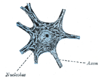
In cellular neuroscience, Nissl bodies (also called Nissl granules, Nissl substance or tigroid substance) are discrete granular structures in neurons that consist of rough endoplasmic reticulum, a collection of parallel, membrane-bound cisternae studded with ribosomes on the cytosolic surface of the membranes.[1] Nissl bodies were named after Franz Nissl, a German neuropathologist who invented the staining method bearing his name (Nissl staining).[2][3] The term "Nissl bodies" generally refers to discrete clumps of rough endoplasmic reticulum and free ribosomes in nerve cells. Masses of rough endoplasmic reticulum also occur in some non-neuronal cells, where they are referred to as ergastoplasm, basophilic bodies,[1] or chromophilic substance.[4] While these organelles differ in some ways from Nissl bodies in neurons,[5] large amounts of rough endoplasmic reticulum are generally linked to the copious production of proteins.[1]
^Richard H. Thompson (29 March 2000). The Brain: A Neuroscience Primer. Macmillan. pp. 35–. ISBN 978-0-7167-3226-6. Retrieved 4 January 2013.
^Da Mota Gomes M (2019). "Franz Nissl (1860-1919), noted neuropsychiatrist and neuropathologist, staining the neuron, but not limiting it". Dementia & Neuropsychologia. 13 (3): 352–355. doi:10.1590/1980-57642018dn13-030014. PMC 6753910.
^Fawcett, Don W. (1966). The Cell: Its Organelles and Inclusions. W.B. Saunders Company. ISBN 0-7216-3585-7.
^Peters A, Palay SL, Webster Hd (1991). The Fine Structure of the Nervous System. Oxford University Press. ISBN 0-19-506571-9.
In cellular neuroscience, Nisslbodies (also called Nissl granules, Nissl substance or tigroid substance) are discrete granular structures in neurons...
Franz Alexander Nissl (9 September 1860, in Frankenthal – 11 August 1919, in Munich) was a German psychiatrist and medical researcher. He was a noted...
Nissl may refer to: Nisslbody, also known as Nissl substance or Nissl material, a component of neurons Franz Nissl, the German medical researcher who...
December 2016. Boeriu, Horatieu (16 January 2014). "Interview with Florian Nissl, BMW M3 and M4 Designer". bmwblog.com. Retrieved 1 September 2022. "A closer...
coloration is due to pyknosis or degradation of the nucleus and loss of Nisslbodies which are normally stained blue (basophilic) on hematoxylin & eosin staining...
February 2008. Boeriu, Horatieu (16 January 2014). "Interview with Florian Nissl, BMW M3 and M4 Designer". bmwblog.com. Retrieved 1 September 2022. Tingwall...
gyri, and even Nisslbodies in the motor cortex could be distinguished in the 20th century. An exposed, insect-infested body or a body in a warm environment...
thionine, cresyl violet, or neutral red. These dyes intensely stain "Nisslbodies" (rough endoplasmic reticulum), which are abundant in neurons and reveal...
cellular neuroscience, chromatolysis is the dissolution of the Nisslbodies in the cell body of a neuron. It is an induced response of the cell usually triggered...
membrane. Numerous microscopic clumps called Nisslbodies (or Nissl substance) are seen when nerve cell bodies are stained with a basophilic ("base-loving")...
Morison Müllerian ducts – Johannes Peter Müller Nisslbodies or granules and Nissl substance – Franz Nissl Sphincter of Oddi – Ruggero Oddi Pacinian corpuscles – Filippo...
Brazil Rudolf Nieuwenhuys 1927– Netherlands Franz Nissl 1860–1919 Germany Nisslbody, Nissl granules, Nissl method Akinori Noma Japan Fernando Nottebohm 1940–...
acidic components of the neuronal cytoplasm a violet colour, specifically nisslbodies. Often used in brain research. Crystal violet, when combined with a suitable...
chromatolysis, the soma and nucleus round and enlarge, the Nisslbodies and Golgi bodies both disintegrate, and the cytocentrum is cleared. In most cases...
telodendria, and axon terminals. It also includes the myelin sheath. The Nisslbodies that produce the neuronal proteins are absent in the axonal region. Proteins...
A motor neuron (or motoneuron or efferent neuron) is a neuron whose cell body is located in the motor cortex, brainstem or the spinal cord, and whose axon...
of the cat superior olive as revealed by PEP-19 immunocytochemistry and Nissl stain". The Journal of Comparative Neurology. 368 (1): 100–120. doi:10...
species of mammals, larger brains (in absolute terms, not just in relation to body size) tend to have thicker cortices. The smallest mammals, such as shrews...
demonstrate iron deposits in diseases like hemochromatosis. The Nissl method for Nissl substance and Golgi's method (and related silver stains) are useful...
used to enhance the contrast of particular features in microscopic images. Nissl staining uses aniline basic dyes to intensely stain the acidic polyribosomes...
neurons are markedly swollen and pear shaped, with the nucleus and the nisslbodies displaced toward the apical dendrites. Examples of neuron metabolic storage...
appearance and location in a neuron and from its sparse distribution of Nissl substance. The axon hillock is the last site in the soma where membrane...
stain Heinz bodies in red blood corpuscles or for staining of the neurons in the brain and spinal cord. It is used to demonstrate the Nissl substance in...
cytoarchitectural organization of neurons he observed in the cerebral cortex using the Nissl method of cell staining. Brodmann published his maps of cortical areas in...
 Global Information