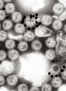
Electron micrograph of gold nanoparticles attached to rotaviruses. The small dark circular objects are gold nanoparticles coated with a monoclonal antibody specific for rotavirus protein VP6.
Immune electron microscopy (more often called immunoelectron microscopy) is the equivalent of immunofluorescence, but it uses electron microscopy rather than light microscopy.[1] Immunoelectron microscopy identifies and localizes a molecule of interest, specifically a protein of interest, by attaching it to a particular antibody. This bond can form before or after embedding the cells into slides. A reaction occurs between the antigen and antibody, causing this label to become visible under the microscope. Scanning electron microscopy is a viable option if the antigen is on the surface of the cell, but transmission electron microscopy may be needed to see the label if the antigen is within the cell.[2]
^"Immuno-Electron Microscopy Services at the Core Electron Microscopy Facility - UMASS Medical School". UMass Chan Medical School. 2 November 2013. Retrieved 5 December 2022.
and 28 Related for: Immune electron microscopy information
Immuneelectronmicroscopy (more often called immunoelectron microscopy) is the equivalent of immunofluorescence, but it uses electronmicroscopy rather...
named coronavirus,: 96 due to their crown-like appearance. Her immuneelectronmicroscopy (IEM) innovations and insights contributed to research related...
his colleagues isolated the Norwalk virus from volunteers using immuneelectronmicroscopy, a process that involves looking directly at antibody-antigen...
(leukocyte) in the immune system of most vertebrates. Lymphocytes include T cells (for cell-mediated and cytotoxic adaptive immunity), B cells (for humoral...
cells (scientific name leukocytes), also called immune cells or immunocytes, are cells of the immune system that are involved in protecting the body against...
The first images of viruses were obtained upon the invention of electronmicroscopy in 1931 by the German engineers Ernst Ruska and Max Knoll. In 1935...
antibody (peroxidase produces a dark brown color).[citation needed] Electronmicroscopy is a method that can take a picture of a whole virus and can reveal...
on bilayers often require advanced techniques like electronmicroscopy and atomic force microscopy. When phospholipids are exposed to water, they self-assemble...
synapses and organelles. Scanning electronmicroscopy, on the other hand, provides 3D information by scanning a focused electron beam across the sample's surface...
stretches and globular domains, as shown via high-resolution scanning electronmicroscopy. After stimulation of the neutrophil response, neutrophils lose their...
children. Although no changes may be visible by light microscopy, changes on electronmicroscopy within the glomeruli may show a fusion of the foot processes...
Transmission electronmicroscopy DNA sequencing is a single-molecule sequencing technology that uses transmission electronmicroscopy techniques. The method...
rotaviruses. Norwalk virus had been discovered by Albert Kapikian using immuneelectronmicroscopy and Ruth Bishop and colleagues had seen different particles that...
century, electronmicroscopy also saw a drastic revolution with the development of more coherent electron sources, aberration correction for electron microscopes...
potential or promised applications in a wide variety of areas, including electronmicroscopy, electronics, nanotechnology, materials science, and biomedicine...
Assembly of new virus particles and release by cell lysis." Using electronmicroscopy, the sapovirus was first seen in diarrheic stool samples from the...
Albert Z.; Purcell, Robert H. (1973). "Hepatitis A: Detection by ImmuneElectronMicroscopy of a Viruslike Antigen Associated with Acute Illness". Science...
proximal tubule of the nephron. Light microscopy findings in LCPT include proximal tubular swelling with electronmicroscopy findings showing proximal tubule...
specific and detect all serotypes of rotavirus. Other methods, such as electronmicroscopy and PCR (polymerase chain reaction), are used in research laboratories...
stain, the GBM appears to have a "spiked" or "holey" appearance. On electronmicroscopy, subepithelial deposits that nestle against the glomerular basement...
Clinical Skin Samples Using Scanning ElectronMicroscopy". In Janecek, Milos; Kral, Robert (eds.). Modern ElectronMicroscopy in Physical and Life Sciences....
organism. Images can be produced from a variety of methods including: microscopy, imaging probes, and spectroscopy. Fluorescence itself, is a form of luminescence...
visualization by fluorescence microscopy and conventional transmission electronmicroscopy (TEM). The resolution of fluorescence microscopy (~200 nm) is insufficient...
during later stages of vessel repair As shown by flow cytometry and electronmicroscopy, the most sensitive sign of activation, when exposed to platelets...
 Global Information