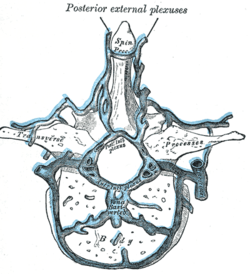
Transverse section of a thoracic vertebra, showing the vertebral venous plexuses.
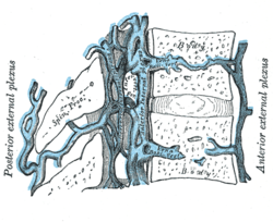
Median sagittal section of two thoracic vertebrae, showing the vertebral venous plexuses.
Details
Identifiers
Latin
plexus venosi vertebrales externi
TA98
A12.3.07.019 A12.3.07.020
TA2
4951, 4952
FMA
12851
Anatomical terminology
[edit on Wikidata]
The external vertebral venous plexuses (extraspinal veins) consist of anterior and posterior plexuses which anastomose freely with each other. They are most prominent in the cervical region[1] where they form anastomoses with the vertebral, occipital, and deep cervical veins.[2]
The anterior external vertebral venous plexuses are situated anteriorly to the vertebral bodies. They communicate with the basivertebral and intervertebral veins, and receive tributaries from the vertebral bodies.
The posterior external vertebral venous plexuses are situated posterior to the vertebral laminae, around and the spinous, transverse, and articular processes. They form anastomoses with the internal vertebral venous plexuses, and drain to vertebral veins, posterior intercostal veins, and lumbar veins.[1]
^ abStandring, Susan (2020). Gray's Anatomy: The Anatomical Basis of Clinical Practice (42nd ed.). New York. p. 882. ISBN 978-0-7020-7707-4. OCLC 1201341621.{{cite book}}: CS1 maint: location missing publisher (link)
^Gray, Henry (1918). Gray's Anatomy (20th ed.). p. 668.
and 30 Related for: External vertebral venous plexuses information
The externalvertebralvenousplexuses (extraspinal veins) consist of anterior and posterior plexuses which anastomose freely with each other. They are...
The internal vertebralvenousplexuses (intraspinal veins) lie within the vertebral canal in the epidural space, embedded within epidural fat. They receive...
suboccipital venousplexus drains deoxygenated blood from the back of the head. It communicates with the externalvertebralvenousplexuses. The external vertebral...
vascular structures slide downwards or when venous pressure is excessively increased. Increased internal and external anal sphincter pressure may also be involved...
that form anastomotic connections between the internal and externalvertebralvenousplexuses may bass between a pair of the ligaments. The ligamenta flava...
also known as the thoracic outlet. There are three main types: neurogenic, venous, and arterial. The neurogenic type is the most common and presents with...
the internal vertebralvenousplexuses into the externalvertebralvenousplexuses. They drain (in craniocaudal sequence) into vertebral vein, intercostal...
pampiniform venousplexus in the scrotum; in a female person, it is an abnormal painful swelling to the embryologically identical pampiniform venousplexus; it...
with the azygos vein (which runs on the right side of the vertebral column) and venousplexuses next to the spinal cord. The inferior vena cava begins as...
The vertebral vein is formed in the suboccipital triangle, from numerous small tributaries which spring from the internal vertebralvenousplexuses and...
vertebralvenousplexuses. Ultimately, this plexus is able to send and receive information from the veins and sinuses inside the brain. The external vertebral...
cerebrospinal venous system (CSVS) consists of the interconnected venous systems of the brain (the cerebral venous system) and the spine (the vertebralvenous system)...
either the brachiocephalic vein, external jugular vein, suprascapular vein, transverse cervical vein, or vertebral vein. In a vast majority of cases...
technique for dissolving blood clots, such as pulmonary embolism and deep venous thrombosis, with either pharmaceutical (TPA) or mechanical means. IVC filters:...
arteriosum Left recurrent laryngeal Azygos Vein Nerves (Cardiac and Pulmonary plexuses) Thoracic duct is a more detailed mnemonic including: Phrenic and Vagus...
the venous and arterial sides of the fetal heart Foramen transversarium, one of a pair of openings in each cervical vertebra, in which the vertebral artery...
behind the diaphragm, at the vertebral level of T12. It travels down the posterior wall of the abdomen, anterior to the vertebral column. It thus follows the...
sacrum, to which it finds attachment. Between the termination of the vertebral column and the anus, the two pubococcygeus muscles come together and form...
mouth, teeth, tongue, and throat. The head rests on the top part of the vertebral column, with the skull joining at C1 (the first cervical vertebra known...
formation of venous spurs and webs. This can lead to narrowing of the vein and cause persistent unilateral leg swelling, contributing to venous thromboembolism...
trapezius and, dipping into the venousplexus of the suboccipital triangle, joins the deep cervical vein and the vertebral vein. Occasionally it follows...
on each side. The lumbar veins communicate with the external and internal vertebralvenousplexuses, and form anastomoses with tributaries of the azygos...
may reverse, with blood draining from the portal venous system, through the plexus. Veins in the plexus may engorge and lead to varices. Esophageal varices...
latter is often further subdivided into the sciatic and pudendal plexuses: The lumbar plexus is formed lateral to the intervertebral foramina by the ventral...
 Global Information