Electrocardiography in myocardial infarction information
Electrocardiography in myocardial infarction
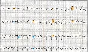
12-lead electrocardiogram showing ST-segment elevation (orange) in I, aVL and V1-V5 with reciprocal changes (blue) in the inferior leads, indicative of an anterior wall myocardial infarction.
Purpose
detecting ischemia or acute coronary injury in emergency department
Electrocardiography in suspected myocardial infarction has the main purpose of detecting ischemia or acute coronary injury in emergency department populations coming for symptoms of myocardial infarction (MI). Also, it can distinguish clinically different types of myocardial infarction.
and 26 Related for: Electrocardiography in myocardial infarction information
Electrocardiographyin suspected myocardialinfarction has the main purpose of detecting ischemia or acute coronary injury in emergency department populations...
representing an occlusive myocardialinfarction (OMI)? Numerous diagnoses and findings can be made based upon electrocardiography, and many are discussed...
cardiovascular diseases. Types include stable angina, unstable angina, and myocardialinfarction. A common symptom is chest pain or discomfort which may travel into...
In medicine, a Holter monitor (often simply Holter) is a type of ambulatory electrocardiography device, a portable device for cardiac monitoring (the...
slightly higher in women than men) and a false negative rate of 20–30%. ST depression may be associated with subendocardial myocardialinfarction, hypokalemia...
ventricular fibrillation unrelated to myocardialinfarction, and 14% of all ventricular fibrillation resuscitations in patients under the age of 40. It follows...
the prognosis. As arrhythmias are relatively common in this group, patients with myocardialinfarction or unstable angina are routinely admitted to the coronary...
Inelectrocardiography, the T wave represents the repolarization of the ventricles. The interval from the beginning of the QRS complex to the apex of the...
breath and associated electrocardiogram (ECG) changes mimicking a myocardialinfarction of the anterior wall. During the course of evaluation of the patient...
department setting focuses primarily on the monitoring of arrhythmia, myocardialinfarction, and QT interval monitoring. It is categorized into one of three...
bradycardia (for example, from drug overdose or myocardialinfarction). A permanent pacemaker may be placed in situations where the bradycardia is not expected...
stenosis (narrowing) in one or more arteries and risking myocardialinfarction, the interruption of blood supply to the heart. CAD can occur in any of the major...
Management of Patients With Unstable Angina/Non-ST-Elevation MyocardialInfarction) developed in collaboration with the American College of Emergency Physicians...
situations in which no cardiac output occurs at all. Without urgent treatment, these events result in sudden death. Myocardialinfarction ("Heart attack")...
is stopped completely, cardiac muscle cells may die, known as a myocardialinfarction, or heart attack. Coronary artery disease (CAD) is the most common...
injury syndromes: Heart surgery (postpericardiotomy syndrome), post-myocardialinfarction (Dressler's syndrome), coronary interventions such as drug eluting...
history of heart disease (e.g. previous myocardialinfarction), as well as heart disease or sudden cardiac death in close relatives. PVCs and palpitation...
Therefore, the length of the pause in heartbeats is usually a multiple of the P-P interval, as seen on electrocardiography. Like a sinus pause, a sinoatrial...
for coronary artery disease and myocardialinfarctions ("heart attacks"). Catheterization is most often performed in special laboratories with fluoroscopy...
bundle branch block. A concordant T wave may suggest ischemia or myocardialinfarction.[citation needed] The underlying condition may be treated by medications...
 Global Information