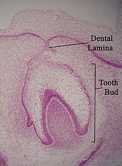
Micrograph of a dental lamina and tooth bud. H&E stain.
Details
Identifiers
Latin
lamina dentalis
TE
lamina_by_E5.4.1.1.1.0.3 E5.4.1.1.1.0.3
Anatomical terminology
[edit on Wikidata]
The dental lamina is a band of epithelial tissue seen in histologic sections of a developing tooth.[1][2] The dental lamina is first evidence of tooth development and begins (in humans) at the sixth week in utero or three weeks after the rupture of the buccopharyngeal membrane. It is formed when cells of the oral ectoderm proliferate faster than cells of other areas. Best described as an in-growth of oral ectoderm, the dental lamina is frequently distinguished from the vestibular lamina, which develops concurrently. This dividing tissue is surrounded by and, some would argue, stimulated by ectomesenchymal growth. When it is present, the dental lamina connects the developing tooth bud to the epithelium of the oral cavity. Eventually, the dental lamina disintegrates into small clusters of epithelium and is resorbed. In situations when the clusters are not resorbed, (this remnant of the dental lamina is sometimes known as the glands of Serres) eruption cysts are formed over the developing tooth and delay its eruption into the oral cavity. This invagination of ectodermal tissues is the progenitor to the later ameloblasts and enamel while the ectomesenchyme[3] is responsible for the dental papilla and later odontoblasts.
^Buchtová, M.; Štembírek, J.; Glocová, K.; Matalová, E.; Tucker, A.S. (2012). "Early Regression of the Dental Lamina Underlies the Development of Diphyodont Dentitions". Journal of Dental Research. 91 (5): 491–498. doi:10.1177/0022034512442896. PMID 22442052. S2CID 206417026.
^Whitlock, John A.; Richman, Joy M. (2013). "Biology of tooth replacement in amniotes". International Journal of Oral Science. 5 (2): 66–70. doi:10.1038/ijos.2013.36. PMC 3707075. PMID 23788284.
^Thesleff, Irma; Tummers, Mark (January 31, 2009). Watt, Fiona; Gage, Fred (eds.). "Tooth organogenesis and regeneration". StemBook. doi:10.3824/stembook.1.37.1. PMID 20614625.
The dentallamina is a band of epithelial tissue seen in histologic sections of a developing tooth. The dentallamina is first evidence of tooth development...
between the vestibular lamina and the dentallamina. It occurs in the sixth to seventh week of the embryonic life. The dentallamina connects the developing...
proliferation of the vestibular lamina into the ectomesenchyme. The vestibular lamina is usually contrasted with the dentallamina, which develops concurrently...
glycogen-rich clear cells, which are also seen in the dentallamina. Therefore, LPC might be related to dentallamina remnants. The epithelial cell rests of Malassez...
strands of dentallamina. The enamel niche is the name of the mesenchymal cells which look to be surrounded by the strands of the dentallamina. In actuality...
Epstein's pearl, is a type of cysts of the jaws that originates from the dentallamina and is found in the mouth parts. It is a superficial cyst in the alveolar...
dental lamina. The dentallamina is a band of epithelial tissue which connects the developing tooth bud to the oral epithelium. The dentallamina eventually...
general dentallamina. Begin to form as early as 24 weeks.[clarification needed] Dental anatomy. ^ Ash, Major M. and Stanley J. Nelson. Wheeler's Dental Anatomy...
reprogrammed to induced pluripotent stem cells which can be placed in the dentallamina directly or placed in a reabsorbable biopolymer in the shape of the...
jaw. The dentallamina is a band of tissue in the developing oral cavity that gives rise to tooth buds. Hyperactivity of the dentallamina, as well as...
developing tooth, including structures known as the enamel organ, dentallamina, and dental papilla. The generally recognized stages of tooth development...
adjacent layer of cuboidal cells (outer enamel epithelium) from the dentallamina. As the cells of the reduced enamel epithelium degenerate, the tooth...
pattern similar to other reptiles where a replacement tooth grows in the dentallamina on the inside of the jaw before migrating outwards, resorbing part of...
In dental anatomy, the lamina limitans is the innermost surface of the dentinal tubule (that exist in dentin) that lies in intimate contact with the long...
usually under or just behind the old tooth, from stem cells in the dentallamina. Young animals typically have a full set of teeth when they hatch; there...
replacement tooth developing from the odontogenic stem cell in the dentallamina. The formation of the teeth is pleurodont; they are fused (ankylosed)...
odontogenic tumor is an odontogenic tumor arising from the enamel organ or dentallamina. Two thirds of cases are located in the anterior maxilla, and one third...
primary teeth starts at the sixth week of tooth development as the dentallamina. This process starts at the midline and then spreads back into the posterior...
(2012). "Early Regression of the DentalLamina Underlies the Development of Diphyodont Dentitions". Journal of Dental Research. 91 (5): 491–498. doi:10...
there is a small replacement tooth and an odontogenic stem cell in the dentallamina in standby that can be activated if required. Crocodilians are more...
tissue, that is, an odontogenic tumor arising from the enamel organ or dentallamina. It may be either truly neoplastic or merely hamartomatous (an odontoma)...
Hermanns syndrome, odontoblastic hyperactivity, mature odontoblasts and dentallamina remnants (Cell Rests of Serres). Gardner's syndrome is a subtype of...
dental extraction (also referred to as tooth extraction, exodontia, exodontics, or informally, tooth pulling) is the removal of teeth from the dental...
focusing on anatomical principle, hypothesised that specific areas of the dentallamina are especially prone to environmental effects during tooth maturation...
Gingival cyst of the newborn; an inclusion cyst from remnants of the dentallamina on a newborn gingiva Gingival cyst of the adult; a soft tissue variant...
 Global Information