Anterior view of the cerebellum. (Tonsil visible at center right.)
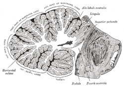
Sagittal section of the cerebellum, near the junction of the vermis with the hemisphere. (Tonsil visible at bottom center.)
Details
Part of
Cerebellum
Artery
PICA
Identifiers
Latin
tonsilla cerebelli
NeuroNames
671
NeuroLex ID
nlx_anat_20081212
TA98
A14.1.07.222
TA2
5817
FMA
83464
Anatomical terms of neuroanatomy
[edit on Wikidata]
The cerebellar tonsil (Latin: tonsilla cerebelli) is a rounded lobule on the undersurface of each cerebellar hemisphere, continuous medially with the uvula of the cerebellar vermis and superiorly by the flocculonodular lobe. Synonyms include: tonsilla cerebelli, amygdala cerebelli, the latter of which is not to be confused with the cerebral tonsils or amygdala nuclei located deep within the medial temporal lobes of the cerebral cortex.
The flocculonodular lobe of the cerebellum, which can also be confused for the cerebellar tonsils, is one of three lobes that make up the overall composition of the cerebellum. The cerebellum consists of three anatomical and functional lobes: anterior lobe, posterior lobe, and flocculonodular lobe.
The cerebellar tonsil is part of the posterior lobe, also known as the neocerebellum, which is responsible for coordinating the voluntary movement of the distal parts of limbs.[1]
Elongation of the cerebellar tonsils can, due to pressure, lead to this portion of the cerebellum to slip or be pushed through the foramen magnum of the skull resulting in tonsillar herniation. This is a life-threatening condition as it causes increased pressure on the medulla oblongata which contains respiratory and cardiac control centres. A congenital condition of tonsillar herniation of either one or both tonsils is Chiari malformation.
^Mavridis, I (2014). "Gross and neurosurgical anatomy of the cerebellar tonsil". S2CID 16241375. {{cite web}}: Missing or empty |url= (help)
The cerebellartonsil (Latin: tonsilla cerebelli) is a rounded lobule on the undersurface of each cerebellar hemisphere, continuous medially with the uvula...
cerebellum, characterized by a downward displacement of one or both cerebellartonsils through the foramen magnum (the opening at the base of the skull)...
Cerebellar peduncles connect the cerebellum to the brain stem. There are six cerebellar peduncles in total, three on each side: Superior cerebellar peduncle...
The posterior inferior cerebellar artery (PICA) is the largest branch of the vertebral artery. It is one of the three main arteries that supply blood to...
herniation, also called downward cerebellar herniation, transforaminal herniation, or "coning", the cerebellartonsils move downward through the foramen...
cause the cerebellartonsil position to descend, which can be mistaken for Chiari malformation; however when the CSF leak is repaired the tonsil position...
malformation of the brain. It consists of a downward displacement of the cerebellartonsils and the medulla through the foramen magnum, sometimes causing hydrocephalus...
tightly folded layer of gray matter, the cerebellar cortex. It has been estimated that if the human cerebellar cortex could be completely unfolded it would...
cord injury is rare. There is also a possibility of herniation of cerebellartonsils when C1/C2 puncture is done laterally. Water-soluble non-ionic iodinated...
Arnold–Chiari malformation, a disorder that takes place when the cerebellartonsils and the medulla oblongata protrude through the foramen magnum into...
cerebellum by the superior cerebellar peduncles, which enter at the caudal end, medially, on the ventral side; the cerebellar peduncles are distinctive...
malformation (CMI) is when the cerebellartonsils push through the foramen magnum of the skull. CSF flow varies based on level of tonsil descent and type of Chiari...
"Pathophysiology of syringomyelia associated with Chiari I malformation of the cerebellartonsils". Journal of Neurosurgery. 80 (1): 3–15. doi:10.3171/jns.1994.80.1...
cover of the hyoglossus muscle and is finally distributed to the palatine tonsil, the mucous membrane of the fauces and base of the tongue, and the serous...
of nerves. accessory nerves. anterior and posterior spinal arteries. the tonsil of the cerebellum (occasionally) as in a tonsillar herniation known as a...
lies above the oral cavity. The adenoids, also known as the pharyngeal tonsils, are lymphoid tissue structures located in the posterior wall of the nasopharynx...
in organ transplant patients). The initial site of infection may be the tonsils, or possibly the gastrointestinal tract. The virus then remains latent...
days later on 22 February. The cause of death was "Herniation of cerebellartonsils and cardio-respiratory arrest due to hemorrhage, brain and eye injuries...
immune individuals, IgA antibodies against poliovirus are present in the tonsils and gastrointestinal tract and able to block virus replication; IgG and...
slaughterhouses, the brain, spinal cord, trigeminal ganglia, intestines, eyes, and tonsils from cattle are classified as specified risk materials, and must be disposed...
however, toxicity and vitamin deficiency can result in the acquired type of cerebellar degeneration disease. Additionally, there are pieces of evidence that...
genes may present as a chronic myelomonocytic leukemia with involvement of tonsil; and d) FGFR1-BCR or FGFR1-MYST3 fusion genes often present with little...
 Global Information