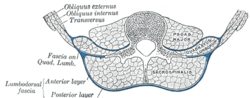
Diagram of a transverse section of the posterior abdominal wall, to show the disposition of the lumbodorsal fascia.
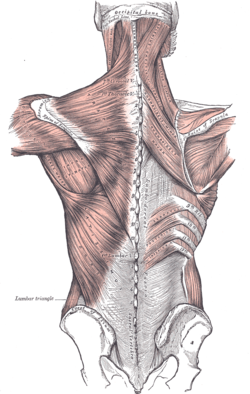
Superficial muscles of the back. The thoracolumbar fascia is the gray area at bottom center.
Details
Identifiers
Latin
fascia thoracolumbalis, fascia lumbodorsalis
TA98
A04.3.02.501
TA2
2242
FMA
25072
Anatomical terminology
[edit on Wikidata]
See also: Lumbar fascia
The thoracolumbar fascia (lumbodorsal fascia or thoracodorsal fascia) is a complex,[1]: 1137 multilayer arrangement of fascial and aponeurotic layers forming a separation between the paraspinal muscles on one side, and the muscles of the posterior abdominal wall (quadratus lumborum, and psoas major[1]: 1137 ) on the other.[2][1]: 1137 It spans the length of the back, extending between the neck superiorly and the sacrum inferiorly.[3] It entails the fasciae and aponeuroses of the latissimus dorsi muscle, serratus posterior inferior muscle, abdominal internal oblique muscle, and transverse abdominal muscle.[4]
In the lumbar region, it is known as lumbar fascia and here consists of 3 layers (posterior, middle, and anterior) enclosing two muscular compartments. In the thoracic region, it consists of a single layer (an upward extension of the posterior layer of the lumbar fascia).[3] The thoracolumbar fascia is most prominent at its lower end[1]: 814–815 where its various layers fuse into a thick composite.[2]
^ abcdStandring S (2020). Gray's Anatomy: The Anatomical Basis of Clinical Practice (42nd ed.). New York. ISBN 978-0-7020-7707-4. OCLC 1201341621.{{cite book}}: CS1 maint: location missing publisher (link)
^ abWillard FH, Vleeming A, Schuenke MD, Danneels L, Schleip R (December 2012). "The thoracolumbar fascia: anatomy, function and clinical considerations". Journal of Anatomy. 221 (6): 507–536. doi:10.1111/j.1469-7580.2012.01511.x. PMC 3512278. PMID 22630613.
^ abSinnatamby C (2011). Last's Anatomy (12th ed.). Elsevier Australia. p. 274. ISBN 978-0-7295-3752-0.
The thoracolumbarfascia (lumbodorsal fascia or thoracodorsal fascia) is a complex,: 1137 multilayer arrangement of fascial and aponeurotic layers forming...
The lumbar fascia is the lumbar portion of the thoracolumbarfascia. It consists of three fascial layers - posterior, middle, and anterior - that enclose...
blends with the psoas fascia and - over the quadratus lumborum muscle - with the anterior layer of thoracolumbarfascia. The iliac fascia overlies the femoral...
with the diaphragm, and from the thoracolumbarfascia. It ends anteriorly in a broad aponeurosis (the Spigelian fascia), the lower fibers of which curve...
Palatine aponeurosis Fascia Willard FH, Vleeming A, Schuenke MD, Danneels L, Schleip R (December 2012). "The thoracolumbarfascia: anatomy, function and...
part of the neck), medially by the serratus anterior muscle and thoracolumbarfascia, anteriorly by the pectoral muscles and posteriorly by the subscapularis...
nuchal fascia is a fascia covering the autochthonous musculature of the neck as a part of the cervical fascia. It proceeds the thoracolumbarfascia to the...
surface of the tendon of the pectoralis major, the coracobrachialis, or the fascia over the biceps brachii. This axillary arch crosses the axillary artery...
underlying transverse fascia. It originates from the inguinal ligament, costal cartilages 7-12, the iliac crest and thoracolumbarfascia. Inserts into the...
layers of muscles, then traverse osteofibrous tunnels between the thoracolumbarfascia and iliac crest. Dysfunction of the superior cluneal nerves is often...
erector spinae is covered in the lumbar and thoracic regions by the thoracolumbarfascia, and in the cervical region by the nuchal ligament. This large muscular...
muscle has a common origin from the iliac crest, the sacrum, the thoracolumbarfascia, and the spinous processes of the vertebrae from T11 to L5. Iliocostalis...
Multifidus Semispinalis Rotatores Interspinales Intertransversarii Thoracolumbarfascia Muscles of thorax Pectoralis major Pectoralis minor Subclavius Serratus...
the iliac crest. It can be thought of as the lower border of the thoracolumbarfascia and is occasionally accompanied by a smaller ligamentous band passing...
irritated upon contact. The superior cluneal nerves travel through the thoracolumbarfascia and drape over the iliac crest. The posterior branches of the iliohypogastric...
veins. The superficial surface of the rib cage is covered by the thoracolumbarfascia, which provides external attachments for the neck, back, pectoral...
between the ilium and the base of the sacrum that is filled by the thoracolumbarfascia and associated muscles. It is generally considered part of the abdominal...
run perpendicular to the external oblique muscle, beginning in the thoracolumbarfascia of the lower back, the anterior 2/3 of the iliac crest (upper part...
angles. The thin aponeurosis of origin is intimately blended with the thoracolumbarfascia, and aponeurosis of the latissimus dorsi muscle.[citation needed]...
represents the thickened inferior border of anterior and middle layers of thoracolumbarfascia. Inferiorly, the ligament is partially continuous with the lumbosacral...
vertebrae. Latissimus dorsi: originates at the supraspinous ligament & thoracolumbarfascia, inserts in the humerus. Antagonist to brachiocephalicus. Supports...
ventral branches of the spinal nerves. Latissimus dorsi: originates on thoracolumbarfascia and inserts on the teres major tuberosity of the humerus. Its function...
J. (2011-01-01), Ross, Mike W.; Dyson, Sue J. (eds.), "Chapter 52 - Thoracolumbar Spine", Diagnosis and Management of Lameness in the Horse (Second Edition)...
specific areas can have benefits such as preventing a postoperative ileus (thoracolumbar junction). Foundations for Osteopathic Medicine by Robert C Ward, et...
ratio AND increased arm/height AND no severe scoliosis = 1 Scoliosis or thoracolumbar kyphosis = 1 Reduced elbow extension = 1 Facial features (3/5) = 1 (dolichocephaly...
 Global Information