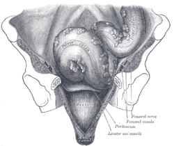
Iliac colon, sigmoid or pelvic colon, and rectum seen from the front, after removal of pubic bones and bladder. (Taenia coli not labeled, but visible at center.)
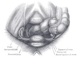
Female pelvis and its contents, seen from above and in front. (Taenia coli not labeled, but visible at right.)
Details
Identifiers
Latin
taeniae coli
TA98
A05.7.03.013
TA2
2993
FMA
76487
Anatomical terminology
[edit on Wikidata]
The taeniae coli (also teniae coli or tenia coli) are three separate longitudinal ribbons (taeniae meaning ribbon in Latin) of smooth muscle on the outside of the ascending, transverse, descending and sigmoid colons. They are visible and can be seen just below the serosa or fibrosa. There are three teniae coli: mesocolic, free and omental taeniae coli. The teniae coli contract lengthwise to produce the haustra, the bulges in the colon.
Large bowel (sigmoid colon) with multiple diverticula. These appear on either side of the longitudinal muscle bundle (taenium).
The bands converge at the root of the vermiform appendix. At the rectosigmoid junction, the taeniae spread out and unite to form the longitudinal muscle layer. In the caecum, the ascending colon, the descending colon and sigmoid colon the positions of these bands are fixed. The taenia libera, is placed anteriorly in the caecum, ascending, descending and sigmoid colon, but is placed inferiorly in the transverse colon. The taenia mesocolica is present on the posteromedial surface of the caecum, ascending, descending and sigmoid colon, but is placed posteriorly on transverse colon at the site of attachment of transverse mesocolon. The taenia omentalis is situated posterolaterally in caecum, ascending, descending and sigmoid colon, but is situated on the anterosuperior surface of transverse colon where layers three and four of the greater omentum meet the transverse colon. This change in position is due to the twist in transverse colon. These bands correspond to the outer layer of the muscularis externa, in other portions of the digestive tract.
The teniae coli are regulated by the sacral nerves of the spinal cord, which are under control of the parasympathetic nervous system.[1]
^Lambert, H. Wayne; Wineski, Lawrence E. (2011). Anatomy & Embryology. Lippincott Williams & Wilkins. p. 65. Destruction of the sacral spinal cord will eliminate parasympathetic outflow to the hindgut, pelvic organs, and perineum as well as somatic innervation to much of the pelvis and lower limbs. Because it stimulates gut motility and tone, loss of parasympathetic input will result in relaxation and inactivity of the teniæ coli in the descending colon.
The taeniae coli (also teniae coli or tenia coli) are three separate longitudinal ribbons (taeniae meaning ribbon in Latin) of smooth muscle on the outside...
polyposis coli, a protein that in humans is encoded by the APC gene Melanosis coli, a disorder of pigmentation of the wall of the colon Taeniacoli, three...
or 'ribbon') may refer to: Taeniacoli, three separate longitudinal ribbons of smooth muscle of the large intestine Taenia thalami, a superior surface...
fat accumulations on the viscera The taeniacoli run the length of the large intestine. Because the taeniacoli are shorter than the large bowel itself...
host cell. This is seen in infections caused by EPEC subgroup Escherichia coli, in celiac disease, and microvillus inclusion disease (an inherited disease...
Unlike other portions of the colon, the rectum does not have distinct taeniae coli. The taeniae blend with one another in the sigmoid colon five centimeters...
colon thins out into 3 discontinuous longitudinal bands, known as taeniae coli (bands of the colon). This is one of the 3 features helping to distinguish...
 Global Information