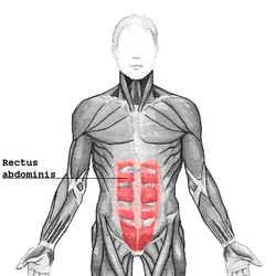
The rectus sheath (also called the rectus fascia[1]) is a tough fibrous compartment formed by the aponeuroses of the transverse abdominal muscle, and the internal and external oblique muscles. It contains the rectus abdominis and pyramidalis muscles, as well as vessels and nerves.[2]
^Te Linde, Richard W. (1977), Rock, John A.; Jones Howard W. (eds.), Te Linde's Operative Gynecology(PDF) (10th ed.), Philadelphia, PA: Lippincott (published 2003), p. 107, retrieved 2018-10-01.
^Sevensma, Karlin E.; Leavitt, Logan; Pihl, Kerent D. (2023), "Anatomy, Abdomen and Pelvis, Rectus Sheath", StatPearls, Treasure Island (FL): StatPearls Publishing, PMID 30725838, retrieved 2023-05-16
oblique muscles. It contains the rectus abdominis and pyramidalis muscles, as well as vessels and nerves. The rectussheath extends between the inferior costal...
The rectus abdominis muscle is contained in the rectussheath, which consists of the aponeuroses of the lateral abdominal muscles. Each rectus abdominus...
posterior layer of the rectussheath inferior to which only the anterior layer of the rectussheath is present and the rectus abdominis muscle is therefore...
A rectussheath hematoma is an accumulation of blood in the sheath of the rectus abdominis muscle. It causes abdominal pain with or without a mass. The...
Diastasis recti, or rectus abdominis diastasis, is defined as a gap of about 2.7 cm or greater between the two sides of the rectus abdominis muscle. The...
Preputial sheath, protective skin around the penis or clitoris Clitoral sheath Penile sheath, the foreskin into which a penis retracts Rectussheath, the laminas...
span more than half the rectus muscle width. It is advisable not to separate the rectus muscles from the anterior rectussheath to prevent their retraction...
arcuate line of rectussheath to enter the rectussheath,: 234 then anastomoses with the superior epigastric artery within the rectussheath.: 225 The inferior...
abdominal wall, and upper rectus abdominis muscle. It enters the rectussheath to descend upon the inner surface of the rectus abdominis muscle. It ends...
defined by the following structures: Medial border: Lateral margin of the rectussheath. Superolateral border: Inferior epigastric vessels. Inferior border:...
is a small triangular muscle, anterior to the rectus abdominis muscle, and contained in the rectussheath. The pyramidalis muscle is part of the anterior...
incision. The rectus fascia and muscle are cut transversely and the incision is extended as far laterally as needed. The anterior rectussheath is not separated...
the rectussheath of the abdominal wall because it is non-absorbable in nature and provides the sheath the due strength it deserves (rectussheath is composed...
insertions such as the pectoral fascia, lower ribs, costal cartilages, rectussheath, aponeurosis of the abdominal external oblique muscle. There is still...
intersections define the anatomy of the rectus abdominis and assist with physiological movement. If the rectus abdominis did not have tendinous intersections...
umbilical fold ends where the vessels reach and enter the rectussheath at the arcuate line of rectussheath; in spite of the name, the lateral umbilical folds...
Perichondral hematoma (ear) Perianal hematoma (anus) Subungual hematoma (nail) Rectussheath hematoma Petechiae – small pinpoint hematomas less than 3 mm in diameter...
band that is made of the bilateral rectussheaths that join at the anterior midline of the body. These enclose the rectus abdominis muscles (a pair of long...
the Rectussheath" Anatomy figure: 35:04-07 at Human Anatomy Online, SUNY Downstate Medical Center - "Incisions and the contents of the rectussheath."...
Fascia The rectussheath (extensive vertical darker gray at left), an example of a fascia Details Precursor Mesenchyme Identifiers Latin fascia MeSH D005205...
prepuce in other mammals is also called the urogenital sinus and clitoral sheath respectively. Vulva hand sign used as a yogic mudra Attic red-figure lid...
approximately the midclavicular line and form the anterior layer of the rectussheath. This aponeurosis formed from fibres from either side of the external...
enter the rectussheath. They run inferiorly, coursing superficially to the fibrous layer forming the posterior leaflet of the rectussheath, and deep...
 Global Information