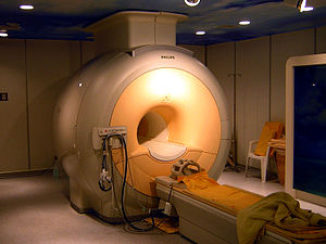
Magnetic resonance imaging (MRI) is a medical imaging technique mostly used in radiology and nuclear medicine in order to investigate the anatomy and physiology of the body, and to detect pathologies including tumors, inflammation, neurological conditions such as stroke, disorders of muscles and joints, and abnormalities in the heart and blood vessels among others. Contrast agents may be injected intravenously or into a joint to enhance the image and facilitate diagnosis. Unlike CT and X-ray, MRI uses no ionizing radiation and is, therefore, a safe procedure suitable for diagnosis in children and repeated runs. Patients with specific non-ferromagnetic metal implants, cochlear implants, and cardiac pacemakers nowadays may also have an MRI in spite of effects of the strong magnetic fields. This does not apply on older devices, and details for medical professionals are provided by the device's manufacturer.
Certain atomic nuclei are able to absorb and emit radio frequency energy when placed in an external magnetic field. In clinical and research MRI, hydrogen atoms are most often used to generate a detectable radio-frequency signal that is received by antennas close to the anatomy being examined. Hydrogen atoms are naturally abundant in people and other biological organisms, particularly in water and fat. For this reason, most MRI scans essentially map the location of water and fat in the body. Pulses of radio waves excite the nuclear spin energy transition, and magnetic field gradients localize the signal in space. By varying the parameters of the pulse sequence, different contrasts may be generated between tissues based on the relaxation properties of the hydrogen atoms therein.
When inside the magnetic field (B0) of the scanner, the magnetic moments of the protons align to be either parallel or anti-parallel to the direction of the field. While each individual proton can only have one of two alignments, the collection of protons appear to behave as though they can have any alignment. Most protons align parallel to B0 as this is a lower energy state. A radio frequency pulse is then applied, which can excite protons from parallel to anti-parallel alignment, only the latter are relevant to the rest of the discussion. In response to the force bringing them back to their equilibrium orientation, the protons undergo a rotating motion (precession), much like a spun wheel under the effect of gravity. The protons will return to the low energy state by the process of spin-lattice relaxation. This appears as a magnetic flux, which yields a changing voltage in the receiver coils to give a signal. The frequency at which a proton or group of protons in a voxel resonates depends on the strength of the local magnetic field around the proton or group of protons, a stronger field corresponds to a larger energy difference and higher frequency photons. By applying additional magnetic fields (gradients) that vary linearly over space, specific slices to be imaged can be selected, and an image is obtained by taking the 2-D Fourier transform of the spatial frequencies of the signal (k-space). Due to the magnetic Lorentz force from B0 on the current flowing in the gradient coils, the gradient coils will try to move producing loud knocking sounds, for which patients require hearing protection.
and 24 Related for: Physics of magnetic resonance imaging information
Nobel Prize in Physics in 1944 for his discovery of nuclear magneticresonance, which is used in magneticresonanceimaging. MR imaging was invented by...
magnetic resonanceimaging (MRI) and nuclear magneticresonance spectroscopy (NMRS) technology. It is also being used to develop nuclear magneticresonance quantum...
Magneticresonanceimaging (MRI) is a medical imaging technique used in radiology to form pictures of the anatomy and the physiological processes inside...
Magneticresonanceimaging (MRI) is in general a safe technique, although injuries may occur as a result of failed safety procedures or human error. During...
Cardiac magneticresonanceimaging (cardiac MRI, CMR), also known as cardiovascular MRI, is a magneticresonanceimaging (MRI) technology used for non-invasive...
Portable magneticresonanceimaging (MRI) is referred to the imaging provided by an MRI scanner that has mobility and portability. It provides MR imaging to...
Magneticresonance microscopy (MRM, μMRI) is magneticresonanceimaging (MRI) at a microscopic level down to the scale of microns. The first definition...
crystals Geophysics Materials physics Medical physics Health physics Radiation dosimetry Medical imagingMagneticresonanceimaging Radiation therapy Microscopy...
In vivo magneticresonance spectroscopy (MRS) is a specialized technique associated with magneticresonanceimaging (MRI). Conventional imaging (lumbar...
techniques, such as in magnetic resonanceimaging (MRI). The original application of NMR to condensed matter physics is nowadays mostly devoted to strongly...
Magneticresonance elastography (MRE) is a form of elastography that specifically leverages MRI to quantify and subsequently map the mechanical properties...
Nuclear magneticresonance spectroscopy, most commonly known as NMR spectroscopy or magneticresonance spectroscopy (MRS), is a spectroscopic technique...
Medical imaging is the technique and process ofimaging the interior of a body for clinical analysis and medical intervention, as well as visual representation...
In physics, optically detected magneticresonance (ODMR) is a double resonance technique by which the electron spin state of a crystal defect may be optically...
biological imaging, digital image restoration, digital imaging, color science, digital photography, holography, magneticresonanceimaging, medical imaging, microdensitometry...
resonances (SR) are a set of spectrum peaks in the extremely low frequency portion of the Earth's electromagnetic field spectrum. Schumann resonances...
Diffusion-weighted magneticresonanceimaging (DWI or DW-MRI) is the use of specific MRI sequences as well as software that generates images from the resulting...
basis of several spectroscopic techniques that are used in condensed matter physics Electron spin resonance Mössbauer effect Nuclear magneticresonance Resonance...
Lauterbur, for discoveries concerning MagneticResonanceImaging (MRI). Mansfield was a professor at the University of Nottingham. Mansfield was born in Lambeth...
electromagnetic radiation. Modern nuclear magneticresonance (NMR) and magneticresonanceimaging (MRI) make use of this effect. The NMR signal observed following...
on the phenomenon of nuclear magneticresonance and adapts a medical magneticresonanceimaging system for the analysis of technical flows. The velocities...
sequence in magneticresonanceimaging (MRI) is a particular setting of pulse sequences and pulsed field gradients, resulting in a particular image appearance...
 Global Information