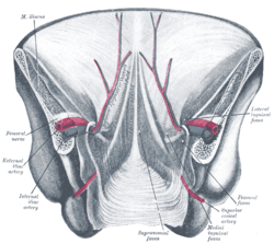
Posterior view of the anterior abdominal wall in its lower half. The peritoneum is in place, and the various cords are shining through. Median umbilical ligament is not labeled, but it is located just underneath the median umbilical fold, seen in the center of the diagram
Not to be confused with Medial umbilical ligament.
In human anatomy, the median umbilical ligament is an unpaired midline ligamentous structure upon the lower inner surface of the anterior abdominal wall.[1] It is covered by the median umbilical fold.[citation needed]
The median umbilical ligament represents the remnant of the fetal urachus.[1][2] It extends from the apex of the bladder to the umbilicus, on the deep surface of the anterior abdominal wall.[2]
The median umbilical ligament represents one of the five ligaments of the internal anterior abdominal wall inferior to the umbilicus; laterally on either side of it are one medial umbilical ligament and finally one lateral umbilical ligament.[1]
^ abcFlynn, William; Vickerton, Paula (2022), "Anatomy, Abdomen and Pelvis, Abdominal Wall", StatPearls, Treasure Island (FL): StatPearls Publishing, PMID 31869113, retrieved 2022-12-17
^ abWilson, Anthony L.; Gandhi, Jason; Seyam, Omar; Rahmani, Benjamin; Patel, Shrey; Joshi, Gunjan; Smith, Noel L.; Khan, Sardar Ali (2019-09-01). "Urachal anomalies: A review of pathological conditions, diagnosis, and management". Translational Research in Anatomy. 16: 100041. doi:10.1016/j.tria.2019.100041. ISSN 2214-854X.
and 30 Related for: Median umbilical ligament information
either side of it are one medial umbilicalligament and finally one lateral umbilicalligament. The medianumbilicalligament begins as the allantois in the...
The medial umbilicalligament, cord of umbilical artery, or obliterated umbilical artery is a paired structure found in human anatomy. It is on the deep...
one medianumbilical fold on the medianumbilicalligament (which in turn, contains the urachus) two medial umbilical folds on the occluded umbilical artery...
needed] Each lateral umbilical fold is situated lateral to the ipsilateral medial umbilical fold. Unlike the median and medial umbilical folds, the contents...
portion obliterates to become the medial umbilicalligament (not to be confused with the medianumbilicalligament, a different structure that represents...
the obliterated umbilical vein, hematogenous spread, or via remnant structures such as the falciform ligament, medianumbilicalligament, or a remnant of...
midline medianumbilicalligament lies medially to each medial umbilical fold; a lateral umbilical fold lies lateral to either medial umbilical fold. A...
1016/j.mpsur.2009.03.002. "Morphologic Variations of the Umbilical Ring, UmbilicalLigaments and Ligamentum Teres Hepatis". ResearchGate. Retrieved 2019-09-02...
The umbilical vein is a vein present during fetal development that carries oxygenated blood from the placenta into the growing fetus. The umbilical vein...
the yolk sac. Normally, the urachus closes off to become the medianumbilicalligament; however, if it does not seal close to the bladder, a blind pouch...
as the medianumbilicalligament. The mouse allantois consists of mesodermal tissue, which undergoes vasculogenesis to form the mature umbilical artery...
canal, the urachus, which later is obliterated and becomes the medianumbilicalligament of the adult. The prostate originally consists of two separate...
from the umbilical plane to the left ribcage. This is the left anterior quadrant in other animals. The right upper quadrant extends from umbilical plane...
later closes as the urachus goes on to definitively form the medianumbilicalligament. Failure of the inside of the urachus to be filled in leaves the...
fetal bladder. After birth the urachus is closed, and becomes the medianumbilicalligament. The fetal membrane surrounds the fetus during the gestational...
toward the upper part of the pubic symphysis, and from there the medianumbilicalligament continues upward on the back of the anterior abdominal wall to...
canal, the urachus, which later is obliterated and becomes the medianumbilicalligament of the adult. Until about the ninth week of gestational age, the...
through a weakness at the site of passage of the umbilical cord through the abdominal wall. Umbilical hernias in adults are largely acquired, and are more...
passed through the umbilical opening, the two arteries, now termed umbilical, enter the umbilical cord, where they coil around the umbilical vein, and ultimately...
uterus, and the vagina. Peritoneal folds are omentums, mesenteries and ligaments; they connect organs to each other or to the abdominal wall. There are...
the greater pelvis, but not in the lesser pelvis. internal iliac artery median sacral artery ovarian artery sacral plexus splanchnic nerves femoral nerve...
cavity, including ascites, blood and pus, tend to collect in this pouch. Median sagittal section of pelvis, showing arrangement of fasciae The peritoneum...
reflect the bladder (vesico-, -vesical) and uterus (utero-, -uterine). Median sagittal section of female pelvis Rectouterine pouch (pouch of Douglas)...
 Global Information