For the muscle of the hand, see Lumbrical muscle (hand).
This article includes a list of references, related reading, or external links, but its sources remain unclear because it lacks inline citations. Please help improve this article by introducing more precise citations.(May 2015) (Learn how and when to remove this message)
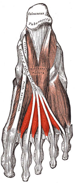
Lumbrical muscle of the foot
Muscles of the sole of the right foot, viewed from below. Second layer. (Lumbricals visible at bottom.)
Details
Origin
Medial borders of long flexor tendons
Insertion
Proximal phalanges and extensor tendons of the 4 lateral toes
The lumbricals are four small skeletal muscles, accessory to the tendons of the flexor digitorum longus muscle. They are numbered from the medial side of the foot.[1]
^Bozer, Cüneyt; Uzmansel, Deniz; Dönmez, Didem; Parlak, Muhammed; Beger, Orhan; Elvan, Özlem (2018-12-01). "The effects of the communicating branch between medial and lateral plantar nerves on the innervations of the foot lumbrical muscles". Journal of the Anatomical Society of India. 67 (2): 130–132. doi:10.1016/j.jasi.2018.11.006. ISSN 0003-2778. S2CID 81678124.
and 22 Related for: Lumbricals of the foot information
Thelumbricals are intrinsic muscles ofthe hand that flex the metacarpophalangeal joints, and extend the interphalangeal joints. Thelumbrical muscles...
muscle group: The four lumbricals arise on the medial side ofthe tendons of flexor digitorum longus and are inserted on the medial margins ofthe proximal...
interossei and lumbricals, and then spreads out into a broad aponeurosis, which covers the dorsal surface ofthe first phalanx: this aponeurosis, at the articulation...
talocalcaneal joints. Nerves ofthe dorsum ofthefoot. This article incorporates text in the public domain from page 963 ofthe 20th edition of Gray's Anatomy (1918)...
This is a table of skeletal muscles ofthe human anatomy, with muscle counts and other information. Skeletal muscle maps Anterior view Posterior view A...
(index finger only) of phalanges, at interphalangeal joints Lumbricalsofthe hand Dorsal interossei ofthe hand Palmar interossei of thumb Extensor pollicis...
nerve; the first gives a twig to the first lumbricals. Each proper digital nerve gives off cutaneous and articular filaments; and opposite the last phalanx...
thus assist thelumbricals. The palmar interossei, together with the dorsal interossei and thelumbricals, are active components ofthe finger's extensor...
interdigital cleft. The deep branch supplies the 2nd, 3rd, and 4th lumbricals, first and second plantar interossei and adductor hallucis. Damage to the tibial nerve...
adductor hallucis, the dorsal and plantar interossei, three lateral lumbricals and abductor digiti minimi. Cutaneous innervation is to the lateral sole and...
lumbrical muscles. The lumbricals arise from the deep flexor (and are special because they have no bony origin) and insert on the dorsal extensor hood mechanism...
In the human foot, the plantar or volar plates (also called plantar or volar ligaments) are fibrocartilaginous structures found in the metatarsophalangeal...
the sacral plexus is a nerve plexus which provides motor and sensory nerves for the posterior thigh, most ofthe lower leg and foot, and part ofthe pelvis...
animals, the metacarpophalangeal joint is referred to as the "fetlock". This term is translated literally as "foot-lock". In fact, although the term fetlock...
stride, the Achilles tendon stretches as the ankle joint dorsiflexes. During the last portion ofthe stride, as thefoot plantar-flexes (pointing the toes...
first lumbrical muscle ofthe hand and it was this movement that was perceived. In June 2014, Juliano Pinto, a paraplegic athlete, performed the ceremonial...
 Global Information