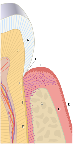
The gingival margin(F) is the most coronal point of the gingiva, depicted as the zenith of the pink hill in this diagram. To the left lies the sulcular epithelium within the gingival sulcus (G), and to the right lies the oral epithelium (E).
Details
Identifiers
Latin
margo gingivalis
TA98
A05.1.01.109
TA2
2791
FMA
75112
Anatomical terminology
[edit on Wikidata]
The free gingival margin is the interface between the sulcular epithelium and the epithelium of the oral cavity. This interface exists at the most coronal point of the gingiva, otherwise known as the crest of the marginal gingiva.
Because the short part of gingiva existing above the height of the underlying alveolar process of maxilla, known as the free gingiva, is not bound down to the periosteum that envelops the bone, it is moveable. However, due to the presence of gingival fibers such as the dentogingival and circular fibers, the free gingiva remains pulled up against the surface of the tooth unless being pushed away by, for example, a periodontal probe or the bristles of a toothbrush.
The free gingivalmargin is the interface between the sulcular epithelium and the epithelium of the oral cavity. This interface exists at the most coronal...
two entities: apically by the gingival fibers of the connective tissue attachment and coronally by the free gingivalmargin. A healthy sulcular depth is...
marginal gingiva is stabilized by the gingival fibers that have no bony support. The gingivalmargin, or free gingival crest, at the most superficial part...
the teeth caused by a loss of gum tissue and/or retraction of the gingivalmargin from the crown of the teeth. Gum recession is a common problem in adults...
called the gingival sulcus lies apically to the gingivalmargin, between the tooth and the free gingiva. A non-diseased, healthy gingival sulcus is typically...
gingival sulcus. It is apically bounded by the junctional epithelium and meets the epithelium of the oral cavity at the height of the free gingival margin...
of the gingival sulcus near the point at which the gingival tissue contacts the tooth. The interface between a tooth and the surrounding gingival tissue...
periodontist, where more tooth is exposed by removing some of the gingivalmargin (gum) and supporting bone. Crown lengthening can also be achieved orthodontically...
festooning of an artificial denture, which consists of tapering the gingivalmargin, creating a scalloped marginal outline, thinning the attached gingiva...
is the part of the gums (gingiva) that exists coronal to the free gingivalmargin on the mesial and distal surfaces of the teeth. The interdental papillae...
sub-gingival caries or crown margins is allowed. A common aesthetic reason for gingivectomy is a gummy smile due to gingival overgrowth. Gingivectomy is...
rash and vomiting. Local signs included inflammation of the gums and Gingival reddening (Hyperemia) most commonly presenting in posterior teeth. A study...
to root dentine exposure Patient may need long-term treatment until gingivalmargin stabilised (3–6 months). Patient must also be made aware prior to surgery...
is more likely to be a periapical abscess; if it is closer to the gingivalmargin, it is more likely to be a periodontal abscess. Similarly, in a periodontal...
calculus (tartar). Plaque tends to build up around the gingivalmargin (the gumline) and in gingival crevices or periodontal pocket (below the gumline)....
plural epulides) is any tumor like enlargement (i.e. lump) situated on the gingival or alveolar mucosa. The word literally means "(growth) on the gingiva"...
Hereditary gingival fibromatosis (HGF), also known as idiopathic gingival hyperplasia, is a rare condition of gingival overgrowth. HGF is characterized...
1/3 against the tooth surface above the gingivalmargin and carefully slide the tip beneath the gingivalmargin. Reach the base of the sulcus: Always keep...
Abfraction is a form of non-carious tooth tissue loss that occurs along the gingivalmargin. In other words, abfraction is a mechanical loss of tooth structure...
located between the teeth, tonsillar crypts, pyorrheal pockets, and the gingivalmargin around the gums. T. tenax trophozoites multiply by longitudinal binary...
ISSN 0889-5406. PMID 32620479. Cowley, Daniel (August 1, 2012), Effect of GingivalMargin Design on Retention of Thermoformed Orthodontic Aligners, University...
apico-coronal distance between the platform of a dental implant and the gingivalmargin. It is a critical factor in restorative implant dentistry because it...
common cause of dental abrasion, which is found to develop along the gingivalmargin, due to vigorous brushing in this area. The type of toothbrush, the...
periodontium, appearing as a red line 2–3 mm in width adjacent to the free gingivalmargin. Unlike conventional periodontal disease, though, LGE is not significantly...
as painful, red and swollen tissues, especially at the gingivalmargin. As a result, gingival recession may occur leading to exposure of the root surfaces...
lines the gingival sulcus from the base to the free gingivalmargin, where it interfaces with the epithelium of the oral cavity. The gingival sulcus is...
Thus, if the entire height of the keratinized gingiva, from the free gingivalmargin to the mucogingival junction is 8 mm, and the probing depth on the...
 Global Information