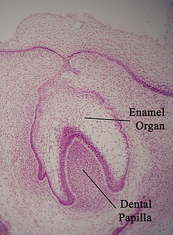
The enamel organ, also known as the dental organ, is a cellular aggregation seen in a developing tooth and it lies above the dental papilla.[1] The enamel organ which is differentiated from the primitive oral epithelium lining the stomodeum. The enamel organ is responsible for the formation of enamel, initiation of dentine formation, establishment of the shape of a tooth's crown, and establishment of the dentoenamel junction.[1]
The enamel organ has four layers; the inner enamel epithelium, outer enamel epithelium, stratum intermedium, and the stellate reticulum.[1]
The dental papilla, the differentiated ectomesenchyme deep to the enamel organ, will produce dentin and the dental pulp. The surrounding ectomesenchyme tissue, the dental follicle, is the primitive cementum, periodontal ligament and alveolar bone beneath the tooth root.[1] The site where the internal enamel epithelium and external enamel epithelium coalesce is the cervical root, important in proliferation of the dental root.[1]
^ abcdeCite error: The named reference Antonio_2018 was invoked but never defined (see the help page).
enamelorgan, also known as the dental organ, is a cellular aggregation seen in a developing tooth and it lies above the dental papilla. The enamel organ...
Tooth enamel is one of the four major tissues that make up the tooth in humans and many animals, including some species of fish. It makes up the normally...
parts: the enamelorgan, the dental papilla and the dental sac or follicle. The enamelorgan is composed of the outer enamel epithelium, inner enamel epithelium...
developing tooth. It lies below a cellular aggregation known as the enamelorgan. The dental papilla appears after 8–10 weeks intra uteral life. The dental...
dental papilla of the enamelorgan in a developing tooth. This layer is first seen during the cap stage, in which these inner enamel epithelium cells are...
the enamelorgan and dental papilla of a developing tooth. It is a vascular fibrous sac containing the developing tooth and its odontogenic organ. The...
The enamel niche is a structure that appears in a histologic slide of a developing tooth from sectioning the slide in a single plane. The enamelorgan looks...
The enamel cord, also called enamel septum, is a localization of cells on an enamelorgan that appear from the outer enamel epithelium to an enamel knot...
parts: the enamelorgan, the dental papilla and the dental follicle. The enamelorgan is composed of the outer enamel epithelium, inner enamel epithelium...
development, the enamel knot is a localization of cells on an enamelorgan that appear thickened in the center of the inner enamel epithelium. The enamel knot is...
stellate reticulum is a group of cells located in the center of the enamelorgan of a developing tooth. These cells are star-shaped and synthesize glycosaminoglycans...
In this defect the enamel is softer than normal. Some areas in enamel are hypocalcified: enamel spindles, enamel tufts, and enamel lamellae. Causal factors...
outer enamel epithelium, also known as the external enamel epithelium, is a layer of cuboidal cells located on the periphery of the enamelorgan in a developing...
invagination of all layers of the enamelorgan in dental papillae. Affected teeth show a deep infolding of enamel and dentin starting from the foramen...
proliferation of epithelial cells located at the cervical loop of the enamelorgan in a developing tooth. Hertwig epithelial root sheath initiates the formation...
cervical loop is the location on an enamelorgan in a developing tooth where the outer enamel epithelium and the inner enamel epithelium join. The cervical...
ameloblastins, enamelins, and tuftelins. The Ca2+ mainly comes from the enamelorgan, and not the dental papilla, by either passive, extracellular transportation...
along with the inner enamel epithelium, is responsible for the tooth enamel formation. It is a part of the dental (enamel) organ. Stratum intermedium...
parts: the enamelorgan, the dental papilla and the dental follicle. The enamelorgan is composed of the outer enamel epithelium, inner enamel epithelium...
adenomatoid odontogenic tumor is an odontogenic tumor arising from the enamelorgan or dental lamina. Two thirds of cases are located in the anterior maxilla...
resulting from invagination of a portion of crown forming within the enamelorgan during odontogenesis. The most extreme form of dens invaginatus is known...
continually produce enamel, they must wear down their teeth by gnawing on various materials. Enamel and dentin are produced by the enamelorgan, and growth is...
Hertwig epithelial root sheath (HERS), near the cervical loop of the enamelorgan. Root dentin is considered different from dentin found in the crown of...
accumulation of fluid either between the reduced enamel epithelium and enamel or in between the layers of enamelorgan seems to be the key to the formation of...
dentin is exposed. In the tooth bud, regions where enamel formation is completed, the enamelorgan gives rise to Hertwig's epithelial root sheath, composed...
ameloblastic tissue, that is, an odontogenic tumor arising from the enamelorgan or dental lamina. It may be either truly neoplastic or merely hamartomatous...
 Global Information