Not to be confused with other types of electrography.
"Micro ERG" redirects here. For the fraction of the unit of energy erg, see micro-erg.
Electroretinography
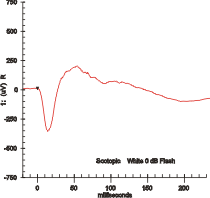
Maximal response ERG waveform from a dark adapted eye.
ICD-9-CM
95.21
MeSH
D004596
[edit on Wikidata]
Schematic Electroretinography waves of healthy people.
Electroretinography measures the electrical responses of various cell types in the retina, including the photoreceptors (rods and cones), inner retinal cells (bipolar and amacrine cells), and the ganglion cells. Electrodes are placed on the surface of the cornea (DTL silver/nylon fiber string or ERG jet) or on the skin beneath the eye (sensor strips) to measure retinal responses. Retinal pigment epithelium (RPE) responses are measured with an EOG test with skin-contact electrodes placed near the canthi. During a recording, the patient's eyes are exposed to standardized stimuli and the resulting signal is displayed showing the time course of the signal's amplitude (voltage). Signals are very small, and typically are measured in microvolts or nanovolts. The ERG is composed of electrical potentials contributed by different cell types within the retina, and the stimulus conditions (flash or pattern stimulus, whether a background light is present, and the colors of the stimulus and background) can elicit stronger response from certain components.[citation needed]
If a dim flash ERG is performed on a dark-adapted eye, the response is primarily from the rod system. Flash ERGs performed on a light adapted eye will reflect the activity of the cone system. Sufficiently bright flashes will elicit ERGs containing an a-wave (initial negative deflection) followed by a b-wave (positive deflection). The leading edge of the a-wave is produced by the photoreceptors, while the remainder of the wave is produced by a mixture of cells including photoreceptors, bipolar, amacrine, and Müller cells or Müller glia.[1] The pattern ERG (PERG), evoked by an alternating checkerboard stimulus, primarily reflects activity of retinal ganglion cells.
^Perlman, Ido. "The Electroretinogram: ERG by Ido Perlman". Webvision at University of Utah. Archived from the original on 2015-12-28.
and 24 Related for: Electroretinography information
Electroretinography measures the electrical responses of various cell types in the retina, including the photoreceptors (rods and cones), inner retinal...
An electrogram (EGM) is a recording of electrical activity of organs such as the brain and heart, measured by monitoring changes in electric potential...
as achromats exhibit a complete absence of cone cell activity via electroretinography in photopic lighting. There are at least four genetic causes of achromatopsia...
088807. PMC 2516064. PMID 18660533. Witzel CA, Joyce JR, Smith EL. Electroretinography of congenital night blindness in an Appaloosa filly. Journal of Equine...
presentation and course. Furthermore, there was greater preservation in electroretinography amplitudes than the more prevalent Pro23His mutation. Autosomal recessive...
Smear tests such as Pap smears dilated fundus examination multifocal electroretinography (mfERG) optical coherence tomography (OCT) visual field test polysomnography...
associated with photopsia, minimal funduscopic changes and abnormal electroretinography findings. This retinal disease was first described by Donald Gass...
angiography to visualize the vascular networks of the retina and choroid. Electroretinography (ERG) measures the electrical responses of various cell types in...
days from post-natal day 6 (p6) to p30 and compared to the vehicle. Electroretinography (ERG), photoreceptor cell counts, cone photoreceptor nuclei counts...
heart Electroencephalography Electrogastrogram Electropalatography Electroretinography Emergency medicine Forward problem of electrocardiology Heart rate...
common electrooculography EOG eye—entire globe 2—somewhat common electroretinography ERG eye—retina specifically 2—somewhat common electronystagmography...
experiments with mice. In humans, evaluation of Surrogate endpoints like electroretinography, auditory evoked potentials and visual analogue scales also suggested...
electroretinography (paracentral depressions) may be obtained. Profound abnormalities detected with visual field and multifocal electroretinography testing...
retinal pathology. Spectral-domain optical coherence tomography, electroretinography and microperimetry are also useful for diagnostic and prognostic...
December 12, 2010. Witzel, C.A.; Joyce, J.R.; Smith, E.L. (1977). "Electroretinography of congenital night blindness in an Appaloosa filly". Journal of...
characteristic chromosomal mutations. An alternative approach is electroretinography, although this is often disfavored for children, since its discomfort...
and the difficulty in making the diagnosis in the early stages, electroretinography (ERG) remains the best test for making the diagnosis. Abnormal cone...
 Global Information