 Global Information
Diffuse optical mammography information
| Diffuse optical mammography | |
|---|---|
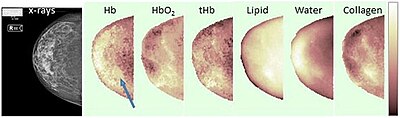 Example of breast constituents' concentrations maps through optical mammography (right cranio-caudal view). The blue arrow points to the lesion. Hb stands for deoxy-hemoglobin, HbO2 for oxy-hemoglobin, tHb for total hemoglobin.[1] | |
| Purpose | investigation of the breast composition through spectral analysis |
Diffuse optical mammography, or simply optical mammography, is an emerging imaging technique that enables the investigation of the breast composition through spectral analysis. It combines in a single non-invasive tool the capability to implement breast cancer risk assessment,[2] lesion characterization,[3] therapy monitoring[4] and prediction of therapy outcome.[5] It is an application of diffuse optics, which studies light propagation in strongly diffusive media, such as biological tissues, working in the red and near-infrared spectral range, between 600 and 1100 nm.[6]
- ^ Cite error: The named reference
Taroni-2017was invoked but never defined (see the help page). - ^ Taroni, Paola; Pifferi, Antonio; Quarto, Giovanna; Spinelli, Lorenzo; Torricelli, Alessandro; Abbate, Francesca; Villa, Anna; Balestreri, Nicola; Menna, Simona; Cassano, Enrico; Cubeddu, Rinaldo (2010). "Noninvasive assessment of breast cancer risk using time-resolved diffuse optical spectroscopy". Journal of Biomedical Optics. 15 (6): 060501–060501–3. Bibcode:2010JBO....15f0501T. doi:10.1117/1.3506043. PMID 21198142.
- ^ Quarto, Giovanna; Spinelli, Lorenzo; Pifferi, Antonio; Torricelli, Alessandro; Cubeddu, Rinaldo; Abbate, Francesca; Balestreri, Nicola; Menna, Simona; Cassano, Enrico; Taroni, Paola (18 September 2014). "Estimate of tissue composition in malignant and benign breast lesions by time-domain optical mammography". Biomedical Optics Express. 5 (10): 3684–3698. doi:10.1364/BOE.5.003684. PMC 4206334. PMID 25360382.
- ^ Jiang, Shudong; Pogue, Brian W.; Carpenter, Colin M.; Poplack, Steven P.; Wells, Wendy A.; Kogel, Christine A.; Forero, Jorge A.; Muffly, Lori S.; Schwartz, Gary N.; Paulsen, Keith D.; Kaufman, Peter A. (August 2009). "Evaluation of Breast Tumor Response to Neoadjuvant Chemotherapy with Tomographic Diffuse Optical Spectroscopy: Case Studies of Tumor Region-of-Interest Changes". Radiology. 252 (2): 551–560. doi:10.1148/radiol.2522081202. PMC 2753781. PMID 19508985.
- ^ Cerussi, A.; Hsiang, D.; Shah, N.; Mehta, R.; Durkin, A.; Butler, J.; Tromberg, B. J. (28 February 2007). "Predicting response to breast cancer neoadjuvant chemotherapy using diffuse optical spectroscopy". Proceedings of the National Academy of Sciences. 104 (10): 4014–4019. Bibcode:2007PNAS..104.4014C. doi:10.1073/pnas.0611058104. PMC 1805697. PMID 17360469.
- ^ Martelli, Fabrizio; Del Bianco, Samuele; Ismaelli, Andrea; Zaccanti, Giovanni (2010). Light propagation through biological tissue and other diffusive media : theory, solutions, and software. SPIE. ISBN 9780819476586.