 Global Information
Codocyte information

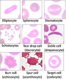
Codocytes, also known as target cells, are red blood cells that have the appearance of a shooting target with a bullseye. In optical microscopy these cells appear to have a dark center (a central, hemoglobinized area) surrounded by a white ring (an area of relative pallor), followed by dark outer (peripheral) second ring containing a band of hemoglobin. However, in electron microscopy they appear very thin and bell shaped (hence the name codo-: bell). Because of their thinness they are referred to as leptocytes. On routine smear morphology, some people like to make a distinction between leptocytes and codocytes- suggesting that in leptocytes the central spot is not completely detached from the peripheral ring, i.e. the pallor is in a C shape rather than a full ring.[1]
These cells are characterized by a disproportional increase in the ratio of surface membrane area to volume. This is also described as a "relative membrane excess." It is due to either increased red cell surface area (increased beyond normal), or else a decreased intracellular hemoglobin content (which may cause an abnormal decrease in cell volume without affecting the amount of membrane area). The increase in the surface area to volume ratio also gives the cell decreased osmotic fragility, as it allows it to take up more water for a given amount of osmotic stress.
In vivo (within the blood vessel), the codocyte is a bell-shaped cell. It assumes a "target" configuration only when processed to obtain a blood film. In the film these cells appear thinner than normal, primarily due to their pallor (by which thickness is judged on microscopy).
When the cells are flattened out on a smear, the top of the bell is pushed to the center creating a central target with a relatively high quantity of hemoglobin.
- ^ Turgeon, Mary Louise (1999). "Ch. 6". Clinical Hematology: Theory and Procedures. Vol. 936. Lippincott Williams & Wilkins. p. 103. ISBN 978-0-316-85623-2.